





.png)


Preparation And Storage


Product Notices
- This reagent has been pre-diluted for use at the recommended Volume per Test. We typically use 1 × 10^6 cells in a 100-µl experimental sample (a test).
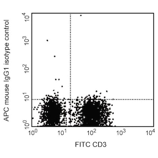
- An isotype control should be used at the same concentration as the antibody of interest.
- Caution: Sodium azide yields highly toxic hydrazoic acid under acidic conditions. Dilute azide compounds in running water before discarding to avoid accumulation of potentially explosive deposits in plumbing.
- Source of all serum proteins is from USDA inspected abattoirs located in the United States.
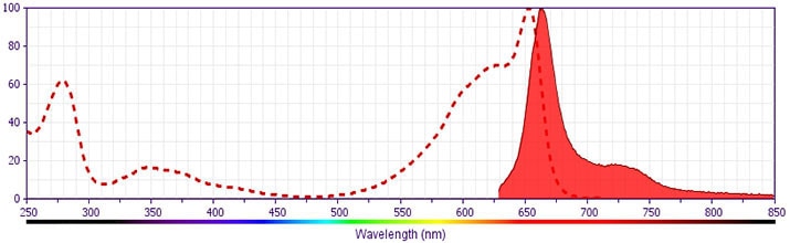
- This APC-conjugated reagent can be used in any flow cytometer equipped with a dye, HeNe, or red diode laser.
- For fluorochrome spectra and suitable instrument settings, please refer to our Multicolor Flow Cytometry web page at www.bdbiosciences.com/colors.
- Please refer to http://regdocs.bd.com to access safety data sheets (SDS).
- Please refer to www.bdbiosciences.com/us/s/resources for technical protocols.
Companion Products


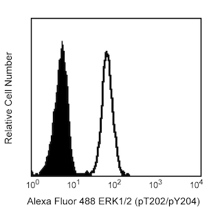

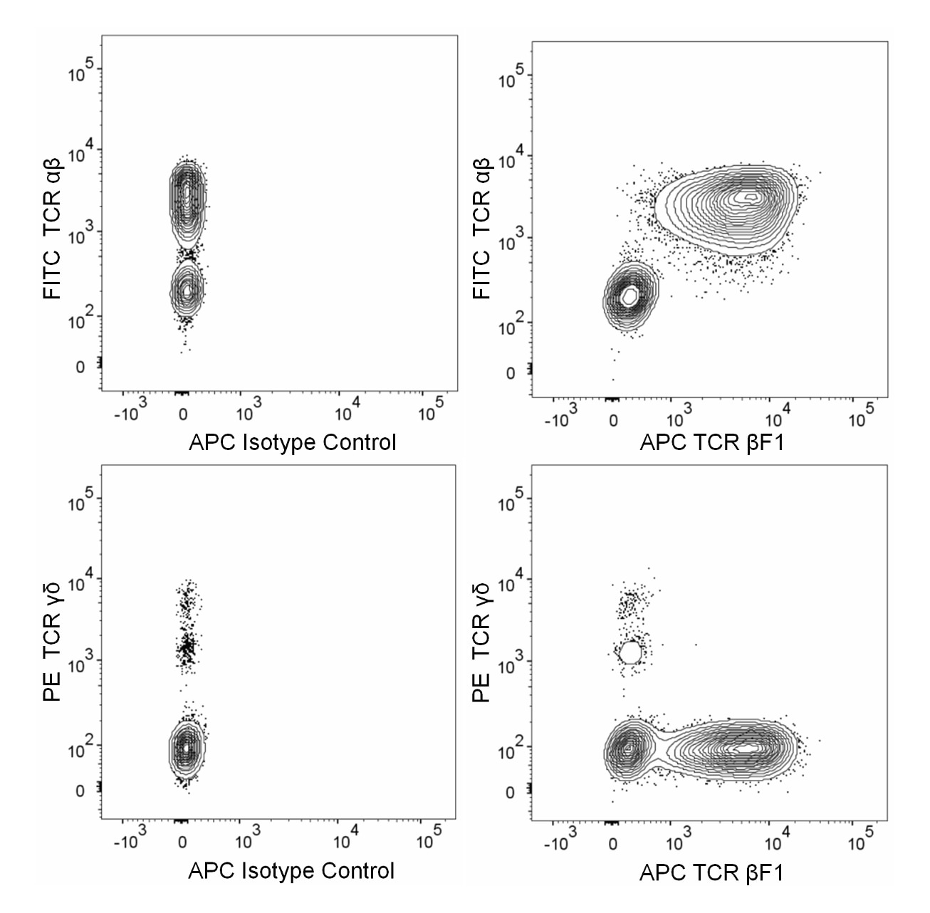
The 8A3 monoclonal antibody is also known as the β Framework 1 (βF1) antibody. After treatment of lymphoid cells with permeabilization reagents, this antibody recognizes a common epitope on the β chain of the T Cell Receptor (TCR) for antigen which is expressed by thymocytes and peripheral T lymphocytes. It does not react with γδ TCR-bearing T cells. The 8A3 antibody does not stain the surface of living cells. This suggests that the TCR β constant region epitope recognized by the 8A3 antibody is normally hidden on viable cells.
Development References (5)
-
Asnafi V, Beldjord K, Boulanger E, et al. Analysis of TCR, pT alpha, and RAG-1 in T-acute lymphoblastic leukemias improves understanding of early human T-lymphoid lineage commitment. Blood. 2003; 101(7):2693-2703. (Clone-specific: Flow cytometry). View Reference
-
Brenner MB, McLean J, Scheft H, Warnke RA, Jones N, Strominger JL. Characterization and expression of the human alpha beta T cell receptor by using a framework monoclonal antibody. J Immunol. 1987; 138(5):1502-1509. (Immunogen: Flow cytometry, Fluorescence microscopy, Immunohistochemistry, Immunoprecipitation, Radioimmunoassay, Western blot). View Reference
-
Fraser JD, Strominger JL. A solubilized T-cell receptor from a human leukemia cell line binds to a ligand in the absence of MHC products.. Immunogenetics. 1988; 28(2):108-16. (Clone-specific: Functional assay). View Reference
-
Ramiro AR, Trigueros C, Márquez C, San Millán JL, Toribio ML. Regulation of pre-T cell receptor (pT alpha-TCR beta) gene expression during human thymic development. J Exp Med. 1996; 184(2):519-530. (Clone-specific: Flow cytometry). View Reference
-
Torres PS, Alcover A, Zapata DA, et al. TCR dynamics in human mature T lymphocytes lacking CD3 gamma. J Immunol. 2003; 170(12):5947-5955. (Clone-specific: Flow cytometry, Fluorescence microscopy, Immunofluorescence). View Reference
Please refer to Support Documents for Quality Certificates
Global - Refer to manufacturer's instructions for use and related User Manuals and Technical data sheets before using this products as described
Comparisons, where applicable, are made against older BD Technology, manual methods or are general performance claims. Comparisons are not made against non-BD technologies, unless otherwise noted.
For Research Use Only. Not for use in diagnostic or therapeutic procedures.
Refer to manufacturer's instructions for use and related User Manuals and Technical Data Sheets before using this product as described.
Comparisons, where applicable, are made against older BD technology, manual methods or are general performance claims. Comparisons are not made against non-BD technologies, unless otherwise noted.
