







Preparation And Storage


Product Notices
- This reagent has been pre-diluted for use at the recommended Volume per Test. We typically use 1 × 10^6 cells in a 100-µl experimental sample (a test).
- An isotype control should be used at the same concentration as the antibody of interest.
- Source of all serum proteins is from USDA inspected abattoirs located in the United States.
- Caution: Sodium azide yields highly toxic hydrazoic acid under acidic conditions. Dilute azide compounds in running water before discarding to avoid accumulation of potentially explosive deposits in plumbing.
- Please observe the following precautions: Absorption of visible light can significantly alter the energy transfer occurring in any tandem fluorochrome conjugate; therefore, we recommend that special precautions be taken (such as wrapping vials, tubes, or racks in aluminum foil) to prevent exposure of conjugated reagents, including cells stained with those reagents, to room illumination.
- For fluorochrome spectra and suitable instrument settings, please refer to our Multicolor Flow Cytometry web page at www.bdbiosciences.com/colors.
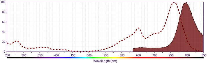
- BD APC-H7 is a tandem conjugate and an analog of APC-Cy7 with the same spectral properties. It has decreased intensity but it is engineered for greater stability and less spillover in the APC channel and consequently offers better performance than APC-Cy7. It has an absorption maximum of approximately 650 nm. When excited by light from a red laser, the APC fluorochrome can transfer energy to the cyanine dye, which then emits at a longer wavelength. The resulting fluorescent emission maximum is approximately 767 nm. BD recommends that a 750-nm longpass filter be used along with a red-sensitive detector such as the Hamamatsu R3896 PMT. As with APC-Cy7 special filters are required when using APC-H7 in conjunction with APC. Note: Although our APC-H7 products demonstrate higher lot-to lot consistency than other APC tandem conjugate products, and every effort is made to minimize the lot-to-lot variation in residual emission from APC, it is strongly recommended that every lot be tested for differences in the amount of compensation required and that individual compensation controls are run for each APC-H7 conjugate.
- Although BD APC-H7 is engineered to minimize spillover to the APC channel and is more stable and less affected by light, temperature, and formaldehyde-based fixatives, compared to other APC-cyanine tandem dyes, it is still good practice to minimize as much as possible, any light, temperature and fixative exposure when working with all fluorescent conjugates.
- Cy is a trademark of GE Healthcare.
- Please refer to www.bdbiosciences.com/us/s/resources for technical protocols.
Companion Products



The H4A3 monoclonal antibody specifically binds to CD107a which is also known as Lysosomal-associated membrane protein 1 (LAMP-1). LAMP-1 is a ~110 kDa type I transmembrane protein that is heavily glycosylated and widely expressed by cells primarily on the luminal surface of their lysosomes. It is also expressed on the surface of activated platelets, activated lymphocytes, cytotoxic T cells and NK cells, and some tumor cell lines, including U937 and KG1a. LAMP-1 can serve as a ligand for E-selectin-mediated cell adhesion. LAMP-1 and LAMP-2 (CD107b) are carriers for poly-N-acetyllactosamines and are able to display sialyl Le[x] termini.
Development References (9)
-
Alto NM, Soderling J, Scott JD. Rab32 is an A-kinase anchoring protein and participates in mitochondrial dynamics. J Biol Chem. 2002; 158(4):659-668. (Biology). View Reference
-
Chen JW, Cha Y, Yuksel KU, Gracy RW, August JT. Isolation and sequencing of a cDNA clone encoding lysosomal membrane glycoprotein mouse LAMP-1. Sequence similarity to proteins bearing onco-differentiation antigens. J Biol Chem. 1988; 263(18):8754-8758. (Biology). View Reference
-
Febbraio M, Silverstein RL. Identification and characterization of LAMP-1 as an activation-dependent platelet surface glycoprotein. J Biol Chem. 1990; 265(30):18531-18537. (Biology). View Reference
-
Fukuda M, Viitala J, Matteson J, Carlsson SR. Cloning of cDNAs encoding human lysosomal membrane glycoproteins, h-lamp-1 and h-lamp-2. Comparison of their deduced amino acid sequences. J Biol Chem. 1988; 263(35):18920-18928. (Biology). View Reference
-
Fukuda M. Lysosomal membrane glycoproteins. Structure, biosynthesis, and intracellular trafficking. J Biol Chem. 1991; 266(32):21327-21330. (Biology). View Reference
-
Hocking DC, Kowalski K. A cryptic fragment from fibronectin's III1 module localizes to lipid rafts and stimulates cell growth and contractility. J Cell Biol. 2002; 158(1):175-184. (Biology). View Reference
-
Sawada R, Lowe JB, Fukuda M. E-selectin-dependent adhesion efficiency of colonic carcinoma cells is increased by genetic manipulation of their cell surface lysosomal membrane glycoprotein-1 expression levels. J Biol Chem. 1993; 268(17):12675-12681. (Biology). View Reference
-
Schlossman SF. Stuart F. Schlossman .. et al., ed. Leucocyte typing V : white cell differentiation antigens : proceedings of the fifth international workshop and conference held in Boston, USA, 3-7 November, 1993. Oxford: Oxford University Press; 1995.
-
Spoerl Z, Stumpf M, Noegel AA, Hasse A. Oligomerization, F-actin interaction, and membrane association of the ubiquitous mammalian coronin 3 are mediated by its carboxyl terminus. J Biol Chem. 2002; 277(50):48858-48867. (Biology). View Reference
Please refer to Support Documents for Quality Certificates
Global - Refer to manufacturer's instructions for use and related User Manuals and Technical data sheets before using this products as described
Comparisons, where applicable, are made against older BD Technology, manual methods or are general performance claims. Comparisons are not made against non-BD technologies, unless otherwise noted.
For Research Use Only. Not for use in diagnostic or therapeutic procedures.
Refer to manufacturer's instructions for use and related User Manuals and Technical Data Sheets before using this product as described.
Comparisons, where applicable, are made against older BD technology, manual methods or are general performance claims. Comparisons are not made against non-BD technologies, unless otherwise noted.
