






.png)


Preparation And Storage


Recommended Assay Procedures
This antibody conjugate is suitable for intracellular staining of human cell lines and peripheral blood mononuclear cells using BD Cytofix™ Fixation Buffer. Any of the three BD Phosflow™ permeabilization buffers may be used.
BD® CompBeads can be used as surrogates to assess fluorescence spillover (Compensation). When fluorochrome conjugated antibodies are bound to BD® CompBeads, they have spectral properties very similar to cells. However, for some fluorochromes there can be small differences in spectral emissions compared to cells, resulting in spillover values that differ when compared to biological controls. It is strongly recommended that when using a reagent for the first time, users compare the spillover on cells and BD® CompBeads to ensure that BD® CompBeads are appropriate for your specific cellular application.
Product Notices
- This reagent has been pre-diluted for use at the recommended Volume per Test. We typically use 1 × 10^6 cells in a 100-µl experimental sample (a test).
- An isotype control should be used at the same concentration as the antibody of interest.
- Source of all serum proteins is from USDA inspected abattoirs located in the United States.
- Caution: Sodium azide yields highly toxic hydrazoic acid under acidic conditions. Dilute azide compounds in running water before discarding to avoid accumulation of potentially explosive deposits in plumbing.
- Alexa Fluor® 488 fluorochrome emission is collected at the same instrument settings as for fluorescein isothiocyanate (FITC).
- For fluorochrome spectra and suitable instrument settings, please refer to our Multicolor Flow Cytometry web page at www.bdbiosciences.com/colors.
- Please refer to www.bdbiosciences.com/us/s/resources for technical protocols.
- This product is provided under an intellectual property license between Life Technologies Corporation and BD Businesses. The purchase of this product conveys to the buyer the non-transferable right to use the purchased amount of the product and components of the product in research conducted by the buyer (whether the buyer is an academic or for-profit entity). The buyer cannot sell or otherwise transfer (a) this product (b) its components or (c) materials made using this product or its components to a third party or otherwise use this product or its components or materials made using this product or its components for Commercial Purposes. Commercial Purposes means any activity by a party for consideration and may include, but is not limited to: (1) use of the product or its components in manufacturing; (2) use of the product or its components to provide a service, information, or data; (3) use of the product or its components for therapeutic, diagnostic or prophylactic purposes; or (4) resale of the product or its components, whether or not such product or its components are resold for use in research. For information on purchasing a license to this product for any other use, contact Life Technologies Corporation, Cell Analysis Business Unit Business Development, 29851 Willow Creek Road, Eugene, OR 97402, USA, Tel: (541) 465-8300. Fax: (541) 335-0504.
- Please refer to http://regdocs.bd.com to access safety data sheets (SDS).
- Alexa Fluor™ is a trademark of Life Technologies Corporation.
Companion Products




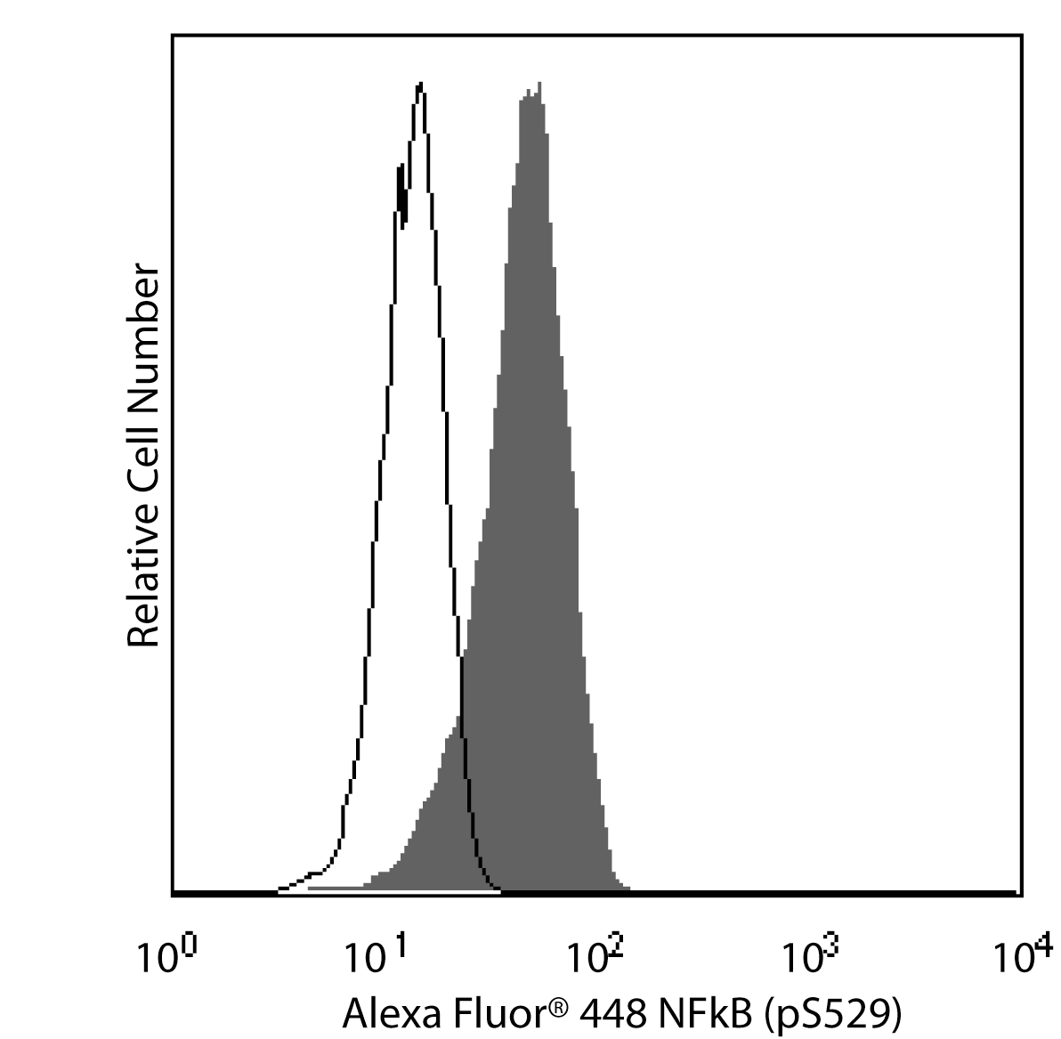
The K10-895.12.50 monoclonal antibody recognizes the phosphorylated serine 529 (pS529) in the transactivation domain of the human NF-κB p65 subunit. Nuclear factor κB (NF-κB) is a ubiquitously expressed transcription factor that regulates the expression of many other genes. It is crucial for cellular responses to a variety of stimuli including stress and microbial pathogens that lead to immunity, inflammation, proliferation, differentiation, survival, apoptosis, and tumorigenesis. The most studied NF-κB complex consists of the p50 (also known as NF-κB1) and p65 (also known as REL-A) subunits, both containing a 300-amino acid region with homology to the Rel proto-oncogene product (RH domain). The RH domain contains motifs for dimerization, nuclear localization, and binding to specific DNA sequences. In addition to the RH domain, the p65 subunit contains the transactivation domain, which is responsible for the interaction with the inhibitor IκB and which contains phosphorylation sites. In most cell types, the p50/p65 heterodimer is located within the cytoplasm complexed to IκB. This complex prevents nuclear translocation and activity of NF-κB. In response to stimuli such as cytokines, LPS, DNA damage, and microbial infections, IκB is phosphorylated at critical residues. This phosphorylation induces dissociation of the IκB/NF-κB complex, allowing the free heterodimeric NF-κB to translocate to the nucleus. Furthermore, optimal activation of NF-κB requires phosphorylation in the transactivation domain of p65. In the nucleus, activated NF-κB dimers bind to the κB sites within promoters and enhancers and function as transcriptional regulators.
Development References (3)
-
Natoli G, Saccani S, Bosisio D, Marazzi I. Interactions of NF-kappaB with chromatin: the art of being at the right place at the right time. Nat Immunol. 2005; 6(5):439-445. (Biology). View Reference
-
Siebenlist U, Brown K, Claudio E. Control of lymphocyte development by nuclear factor-kappaB. Nat Rev Immunol. 2005; 5:435-445. (Biology). View Reference
-
Viatour P, Merville M-P, Bours V, Chariot A. Phosphorylation of NF-kappaB and IkappaB proteins: implications in cancer and inflammation. Trends Biochem Sci. 2005; 30(1):43-52. (Biology). View Reference
Please refer to Support Documents for Quality Certificates
Global - Refer to manufacturer's instructions for use and related User Manuals and Technical data sheets before using this products as described
Comparisons, where applicable, are made against older BD Technology, manual methods or are general performance claims. Comparisons are not made against non-BD technologies, unless otherwise noted.
For Research Use Only. Not for use in diagnostic or therapeutic procedures.
Refer to manufacturer's instructions for use and related User Manuals and Technical Data Sheets before using this product as described.
Comparisons, where applicable, are made against older BD technology, manual methods or are general performance claims. Comparisons are not made against non-BD technologies, unless otherwise noted.
