Skip to main content Skip to navigation
Purified Mouse Anti-Rat CD11b/c
Immunohistochemical staining of granulocytes and macrophages. A frozen section from rat spleen was stained with Mouse Anti-Rat CD11b/c (Cat.550299). Granulocytes and macrophages can be identified by the intense brown labeling of their cell surface membranes (40x magnification).
Product Details
Down Arrow Up Arrow


BD Pharmingen™
Itgam/Integrin, alpha M, C3bi receptor, CR3; Itgad/Integrin, alpha D
Rat (QC Testing)
Mouse BALB/c IgG2a, κ
Resident peritoneal cells from (PVG.RT1[c] x PVG.RT1[u]) and (PVG.RT1[c] x PVG.RT1[a]) F1-hybrid rat
Flow cytometry (Routinely Tested), Immunohistochemistry-frozen, Immunohistochemistry-zinc-fixed (Tested During Development), Immunohistochemistry-formalin (antigen retrieval required) (Not Recommended)
62.5 µg/ml
AB_393594
Aqueous buffered solution containing BSA, goat serum, and ≤0.09% sodium azide.
RUO


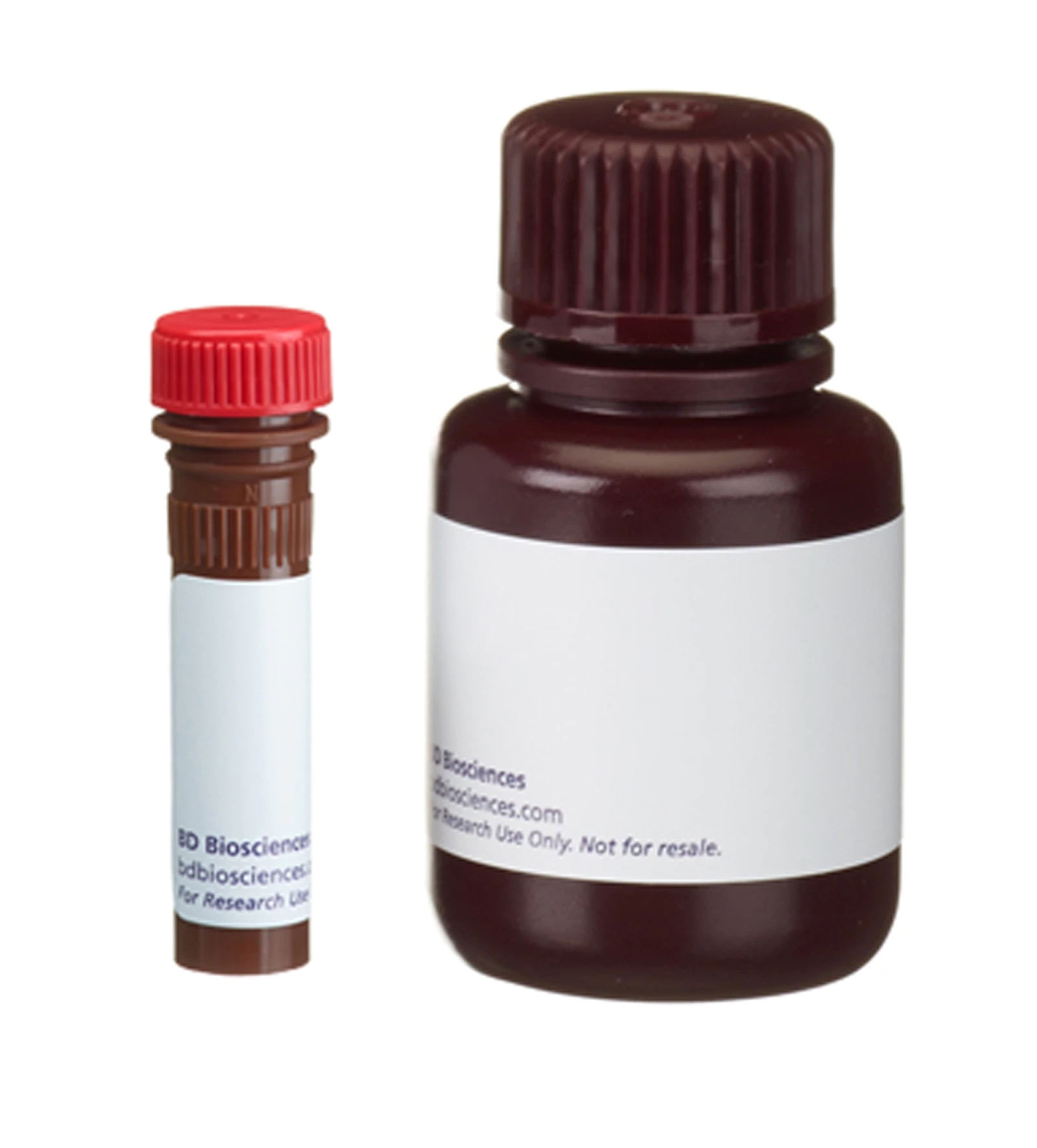
Preparation And Storage

Store undiluted at 4°C. The monoclonal antibody was purified from tissue culture supernatant or ascites by affinity chromatography.

Recommended Assay Procedures

Immunohistochemistry:  This antibody may be used for immunohistochemical staining of acetone-fixed, frozen and zinc-fixed, paraffin sections. This antibody is not recommended for formalin-fixed, paraffin-embedded sections.  This antibody has been reported to stain granulocytes and macrophages in tissues such as rat spleen and thymus. The isotype control suggested for use with this antibody is Purified Mouse IgG2a κ Isotype Control (Cat. No. 550339). For optimal indirect immunohistochemical staining, this antibody should be titrated (1:10 to 1:50 dilution) and visualized via a three-step staining procedure in combination with Biotin Rat Anti-Mouse IgG2a  (Cat. No. 550332) as the secondary antibody and Streptavidin HRP (Cat. No. 550946) together with a DAB Substrate Kit (Cat. No. 550880).  

Product Notices

  1. Since applications vary, each investigator should titrate the reagent to obtain optimal results.
  2. An isotype control should be used at the same concentration as the antibody of interest.
  3. Caution: Sodium azide yields highly toxic hydrazoic acid under acidic conditions. Dilute azide compounds in running water before discarding to avoid accumulation of potentially explosive deposits in plumbing.
  4. Source of all serum proteins is from USDA inspected abattoirs located in the United States.
  5. This antibody has been developed for the immunohistochemistry application. However, a routine immunohistochemistry test is not performed on every lot. Researchers are encouraged to titrate the reagent for optimal performance.
  6. Sodium azide is a reversible inhibitor of oxidative metabolism; therefore, antibody preparations containing this preservative agent must not be used in cell cultures nor injected into animals. Sodium azide may be removed by washing stained cells or plate-bound antibody or dialyzing soluble antibody in sodium azide-free buffer. Since endotoxin may also affect the results of functional studies, we recommend the NA/LE (No Azide/Low Endotoxin) antibody format, if available, for in vitro and in vivo use.
  7. Please refer to www.bdbiosciences.com/us/s/resources for technical protocols.
550299 Rev. 4
Antibody Details
Down Arrow Up Arrow
OX-42

The OX-42 monoclonal antibody specifically binds to the CR3 complement (C3bi) receptor found on most monocytes, granulocytes, macrophages, dendritic cells, and microglia. It appears to recognize a common epitope shared by CD11b and CD11c (integrin αM and αX chains). OX-42 antibody inhibits C3bi binding activity.

550299 Rev. 4
Format Details
Down Arrow Up Arrow
Purified
Tissue culture supernatant is purified by either protein A/G or affinity purification methods. Both methods yield antibody in solution that is free of most other soluble proteins, lipids, etc. This format provides pure antibody that is suitable for a number of downstream applications including: secondary labeling for flow cytometry or microscopy, ELISA, Western blot, etc.
Purified
550299 Rev.4
Citations & References
Down Arrow Up Arrow

Development References (6)

  1. Issekutz AC, Issekutz TB. A major portion of polymorphonuclear leukocyte and T lymphocyte migration to arthritic joints in the rat is via LFA-1/MAC-1-independent mechanisms. Clin Immunol Immunopathol. 1993; 67(3):257-263. (Biology: Blocking). View Reference
  2. Issekutz AC, Issekutz TB. The contribution of LFA-1 (CD11a/CD18) and MAC-1 (CD11b/CD18) to the in vivo migration of polymorphonuclear leucocytes to inflammatory reactions in the rat. Immunology. 1992; 76(4):655-661. (Biology: Blocking). View Reference
  3. Robinson AP, White TM, Mason DW. MRC OX-43: a monoclonal antibody which reacts with all vascular endothelium in the rat except that of brain capillaries. Immunology. 1986; 57(2):231-237. (Immunogen). View Reference
  4. Robinson AP, White TM, Mason DW. Macrophage heterogeneity in the rat as delineated by two monoclonal antibodies MRC OX-41 and MRC OX-42, the latter recognizing complement receptor type 3. Immunology. 1986; 57(2):239-247. (Immunogen: Blocking, Immunoprecipitation). View Reference
  5. Tamatani T, Kotani M, Miyasaka M. Characterization of the rat leukocyte integrin, CD11/CD18, by the use of LFA-1 subunit-specific monoclonal antibodies. Eur J Immunol. 1991; 21(3):627-633. (Biology: Immunoprecipitation). View Reference
  6. Yamazaki T, Seko Y, Tamatani T, et al. Expression of intercellular adhesion molecule-1 in rat heart with ischemia/reperfusion and limitation of infarct size by treatment with antibodies against cell adhesion molecules. Am J Pathol. 1993; 143(2):410-418. (Biology: Blocking). View Reference
View All (6) View Less
550299 Rev. 4

Please refer to Support Documents for Quality Certificates


Global - Refer to manufacturer's instructions for use and related User Manuals and Technical data sheets before using this products as described


Comparisons, where applicable, are made against older BD Technology, manual methods or are general performance claims.  Comparisons are not made against non-BD technologies, unless otherwise noted.

For Research Use Only. Not for use in diagnostic or therapeutic procedures.