-
Your selected country is
Middle East / Africa
- Change country/language
Old Browser
This page has been recently translated and is available in French now.
Looks like you're visiting us from {countryName}.
Would you like to stay on the current country site or be switched to your country?


Two color flow cytometric analysis of CD19 expression on mouse splenocytes. Splenic leucocytes from a BALB/c mouse were preincubated with Purified Rat Anti-Mouse CD16/CD32 antibody (Mouse BD Fc Block™) (Cat. No. 553141/553142). The cells were then stained with APC Hamster Anti-Mouse CD3e antibody (Cat. No. 553066/561826) and either BD Horizon™ BUV395 Rat IgG2a, κ Isotype Control (Cat. No. 563556; Left Panel) or BD Horizon™ BUV395 Rat Anti-Mouse CD19 antibody (Cat. No. 563557/565965; Right Panel). The two-color fluorescence dot plots show the correlated expression patterns of CD19 (or Ig Isotype control staining) versus CD3 for gated events with the forward and side light-scatter characteristic of viable splenic leucocytes. Flow cytometric analysis was performed using a BD™ LSR II Flow Cytometer System.


BD Horizon™ BUV395 Rat Anti-Mouse CD19

Regulatory Status Legend
Any use of products other than the permitted use without the express written authorization of Becton, Dickinson and Company is strictly prohibited.
Preparation And Storage
Recommended Assay Procedures
For optimal and reproducible results, BD Horizon Brilliant™ Stain Buffer should be used anytime BD Horizon Brilliant™ dyes are used in a multicolor flow cytometry panel. Fluorescent dye interactions may cause staining artifacts which may affect data interpretation. The BD Horizon Brilliant Stain Buffer was designed to minimize these interactions. When BD Horizon Brilliant Stain Buffer is used in in the multicolor panel, it should also be used in the corresponding compensation controls for all dyes to achieve the most accurate compensation. For the most accurate compensation, compensation controls created with either cells or beads should be exposed to BD Horizon Brilliant Stain Buffer for the same length of time as the corresponding multicolor panel. More information can be found in the Technical Data Sheet of the BD Horizon Brilliant Stain Buffer (Cat. No. 563794/566349) or the BD Horizon Brilliant Stain Buffer Plus (Cat. No. 566385).
Product Notices
- Since applications vary, each investigator should titrate the reagent to obtain optimal results.
- An isotype control should be used at the same concentration as the antibody of interest.
- Source of all serum proteins is from USDA inspected abattoirs located in the United States.
- Caution: Sodium azide yields highly toxic hydrazoic acid under acidic conditions. Dilute azide compounds in running water before discarding to avoid accumulation of potentially explosive deposits in plumbing.
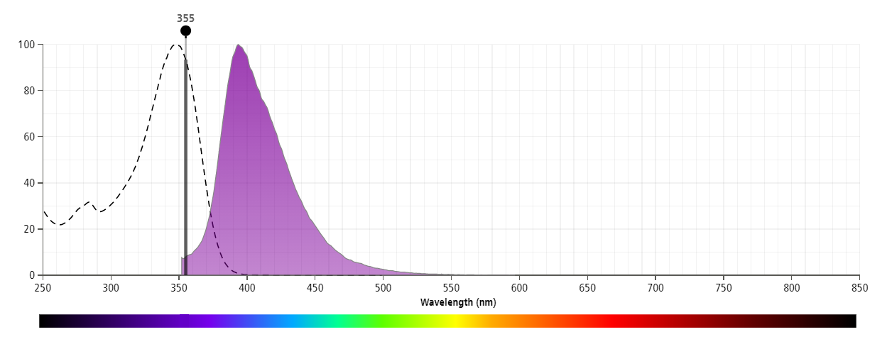
- For fluorochrome spectra and suitable instrument settings, please refer to our Multicolor Flow Cytometry web page at www.bdbiosciences.com/colors.
- BD Horizon Brilliant Ultraviolet 395 is covered by one or more of the following US patents: 8,158,444; 8,575,303; 8,354,239.
- BD Horizon Brilliant Stain Buffer is covered by one or more of the following US patents: 8,110,673; 8,158,444; 8,575,303; 8,354,239.
- Please refer to www.bdbiosciences.com/us/s/resources for technical protocols.
Companion Products


The 1D3 antibody reacts with CD19, a B lymphocyte-lineage differentiation antigen. CD19, a 95-kDa transmembrance glycoprotein, is a member of the immunoglobulin superfamily and is expressed throughout B-lymphocyte development from the pro-B cell through the mature B-cell stages. Terminally differentiated plasma cells do not express CD19. On the surface of mature B cells, the CD19 molecule associates with CD21 (CR-2) and CD81 (TAPA-1), and this multimolecular complex synergizes with surface immunoglobulin to promote cellular activation. Studies with CD19-deficient mice have suggested that the level of CD19 expression affects the generation and maturation of B cells in the bone marrow and periphery. B-1 lineage B cells, also known as CD5+ B cells, are drastically reduced or absent in CD19-deficient mice. Increased levels of CD19 expression correlate with increased frequencies of peritonal and splenic B-1 cells and reduced numbers of conventional B lymphocytes in the periphery. CD19 participates in B-lymphocyte development, B-cell activation, maturation of memory B cells and regulation of tolerance. CD19 has also been detected on peritoneal mast cells, co-localized with CD21/CD35, and it is proposed to play a role in complement-mediated mast-cell activation.

Development References (12)
-
Engel P, Zhou LJ, Ord DC, Sato S, Koller B, Tedder TF. Abnormal B lymphocyte development, activation, and differentiation in mice that lack or overexpress the CD19 signal transduction molecule. Immunity. 1995; 3(1):39-50. (Biology). View Reference
-
Fearon DT. The CD19-CR2-TAPA-1 complex, CD45 and signaling by the antigen receptor of B lymphocytes. Curr Opin Immunol. 1993; 5(3):341-348. (Biology). View Reference
-
Gommerman JL, Oh DY, Zhou X, et al. A role for CD21/CD35 and CD19 in responses to acute septic peritonitis: a potential mechanism for mast cell activation. J Immunol. 2000; 165(12):6915-6921. (Biology). View Reference
-
Inaoki M, Sato S, Weintraub BC, Goodnow CC, Tedder TF. CD19-regulated signaling thresholds control peripheral tolerance and autoantibody production in B lymphocytes. J Exp Med. 1997; 186(11):1923-1931. (Biology). View Reference
-
Krop I, Shaffer AL, Fearon DT, Schlissel MS. The signaling activity of murine CD19 is regulated during cell development. J Immunol. 1996; 157(1):48-56. (Clone-specific: Activation, Calcium Flux, (Co)-stimulation, Flow cytometry, Functional assay, Immunoprecipitation). View Reference
-
Krop I, de Fougerolles AR, Hardy RR, Allison M, Schlissel MS, Fearon DT. Self-renewal of B-1 lymphocytes is dependent on CD19. Eur J Immunol. 1996; 26(1):238-242. (Immunogen: Flow cytometry, Fluorescence activated cell sorting, Functional assay, Immunoprecipitation, In vivo exacerbation). View Reference
-
Rickert RC, Rajewsky K, Roes J. Impairment of T-cell-dependent B-cell responses and B-1 cell development in CD19-deficient mice. Nature. 1995; 376(6538):352-355. (Biology). View Reference
-
Sato S, Jansen PJ, Tedder TF. CD19 and CD22 expression reciprocally regulates tyrosine phosphorylation of Vav protein during B lymphocyte signaling. Proc Natl Acad Sci U S A. 1997; 94(24):13158-13162. (Biology). View Reference
-
Sato S, Miller AS, Howard MC, Tedder TF. Regulation of B lymphocyte development and activation by the CD19/CD21/CD81/Leu 13 complex requires the cytoplasmic domain of CD19. J Immunol. 1997; 159(7):3278-3287. (Biology). View Reference
-
Sato S, Ono N, Steeber DA, Pisetsky DS, Tedder TF. CD19 regulates B lymphocyte signaling thresholds critical for the development of B-1 lineage cells and autoimmunity. J Immunol. 1996; 157(10):4371-4378. (Biology). View Reference
-
Sato S, Steeber DA,Jansen PJ, Tedder TF. CD19 expression levels regulate B lymphocyte development: human CD19 restores normal function in mice lacking endogenous CD19. J Immunol. 1997; 158(10):4662-4669. (Biology). View Reference
-
Tedder TF, Zhou LJ, Engel P. The CD19/CD21 signal transduction complex of B lymphocytes. Immunol Today. 1994; 15(9):437-442. (Biology). View Reference
Please refer to Support Documents for Quality Certificates
Global - Refer to manufacturer's instructions for use and related User Manuals and Technical data sheets before using this products as described
Comparisons, where applicable, are made against older BD Technology, manual methods or are general performance claims. Comparisons are not made against non-BD technologies, unless otherwise noted.
For Research Use Only. Not for use in diagnostic or therapeutic procedures.
Report a Site Issue
This form is intended to help us improve our website experience. For other support, please visit our Contact Us page.