-
Reagents
- Flow Cytometry Reagents
-
Western Blotting and Molecular Reagents
- Immunoassay Reagents
-
Single-Cell Multiomics Reagents
- BD® OMICS-Guard Sample Preservation Buffer
- BD® AbSeq Assay
- BD® Single-Cell Multiplexing Kit
- BD Rhapsody™ ATAC-Seq Assays
- BD Rhapsody™ Whole Transcriptome Analysis (WTA) Amplification Kit
- BD Rhapsody™ TCR/BCR Next Multiomic Assays
- BD Rhapsody™ Targeted mRNA Kits
- BD Rhapsody™ Accessory Kits
- BD® OMICS-One Protein Panels
-
Functional Assays
-
Microscopy and Imaging Reagents
-
Cell Preparation and Separation Reagents
Old Browser
Looks like you're visiting us from {countryName}.
Would you like to stay on the current location site or be switched to your location?
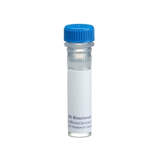
BD Transduction Laboratories™ Purified Mouse Anti-Human CD104
Clone 7/CD104 (RUO)


Western blot analysis of CD104 (Integrin β4) on a A431 cell lysate (Human epithelial carcinoma; ATCC CRL-1555). Lane 1: 1:250, lane 2: 1:500, lane 3: 1:1000 dilution of the anti- human CD104 antibody.

Western blot analysis of CD104 (Integrin β4) on a A431 cell lysate (Human epithelial carcinoma; ATCC CRL-1555). Lane 1: 1:250, lane 2: 1:500, lane 3: 1:1000 dilution of the anti- human CD104 antibody.

Immunofluorescence staining of A431 cells (Human epithelial carcinoma; ATCC CRL-1555).


Regulatory Status Legend
Any use of products other than the permitted use without the express written authorization of Becton, Dickinson and Company is strictly prohibited.
Preparation And Storage
Recommended Assay Procedures
Western blot: Please refer to http://www.bdbiosciences.com/pharmingen/protocols/Western_Blotting.shtml
Product Notices
- Since applications vary, each investigator should titrate the reagent to obtain optimal results.
- Please refer to www.bdbiosciences.com/us/s/resources for technical protocols.
- Caution: Sodium azide yields highly toxic hydrazoic acid under acidic conditions. Dilute azide compounds in running water before discarding to avoid accumulation of potentially explosive deposits in plumbing.
- Source of all serum proteins is from USDA inspected abattoirs located in the United States.
Companion Products


Cell adhesion to extracellular matrix components or to cell surface proteins, especially those expressed by leukocytes and endothelial cells, is mediated by integrins. Integrins contain noncovalently associated α and β subunits. At least 17 α and 8 β subunits have been identified and these proteins can heterodimerize to form 22 different receptors. The α6β4 integrin is a receptor for various laminins and binds with the highest affinity to laminins 4 and 5. It exhibits elevated expression in the basal cell layer of stratified epithelia, in Schwann cells at the onset of myelination, and in CD4-CD8- pre-T lymphocytes entering the thymus. In addition, α6β4 expression is increased in squamous carcinomas where it promotes invasion through a targeting of PI3 kinase activity. The majority of β4 comprises a cytoplasmic domain with unique signaling properties. The C-terminal portion of this domain contains two pairs of type III fibronectin-like motifs (FNIII) and a tyrosine activation motif (TAM). Additional domains in the cytoplasmic tail bind Shc and activate the MAPK pathway. Thus, integrin β4 is an integrin subunit that is important for cell survival, growth, and differentiation.
Development References (2)
-
Mainiero F, Pepe A, Wary KK, et al. Signal transduction by the alpha 6 beta 4 integrin: distinct beta 4 subunit sites mediate recruitment of Shc/Grb2 and association with the cytoskeleton of hemidesmosomes. EMBO J. 1995; 14(18):4470-4481. (Biology). View Reference
-
Shaw LM, Rabinovitz I, Wang HH, Toker A, Mercurio AM. Activation of phosphoinositide 3-OH kinase by the alpha6beta4 integrin promotes carcinoma invasion. Cell. 1997; 91(7):949-960. (Biology). View Reference
Please refer to Support Documents for Quality Certificates
Global - Refer to manufacturer's instructions for use and related User Manuals and Technical data sheets before using this products as described
Comparisons, where applicable, are made against older BD Technology, manual methods or are general performance claims. Comparisons are not made against non-BD technologies, unless otherwise noted.
For Research Use Only. Not for use in diagnostic or therapeutic procedures.
Refer to manufacturer's instructions for use and related User Manuals and Technical Data Sheets before using this product as described.
Comparisons, where applicable, are made against older BD technology, manual methods or are general performance claims. Comparisons are not made against non-BD technologies, unless otherwise noted.