-
Reagents
- Flow Cytometry Reagents
-
Western Blotting and Molecular Reagents
- Immunoassay Reagents
-
Single-Cell Multiomics Reagents
- BD® OMICS-Guard Sample Preservation Buffer
- BD® AbSeq Assay
- BD® Single-Cell Multiplexing Kit
- BD Rhapsody™ ATAC-Seq Assays
- BD Rhapsody™ Whole Transcriptome Analysis (WTA) Amplification Kit
- BD Rhapsody™ TCR/BCR Next Multiomic Assays
- BD Rhapsody™ Targeted mRNA Kits
- BD Rhapsody™ Accessory Kits
- BD® OMICS-One Protein Panels
-
Functional Assays
-
Microscopy and Imaging Reagents
-
Cell Preparation and Separation Reagents
-
- BD® OMICS-Guard Sample Preservation Buffer
- BD® AbSeq Assay
- BD® Single-Cell Multiplexing Kit
- BD Rhapsody™ ATAC-Seq Assays
- BD Rhapsody™ Whole Transcriptome Analysis (WTA) Amplification Kit
- BD Rhapsody™ TCR/BCR Next Multiomic Assays
- BD Rhapsody™ Targeted mRNA Kits
- BD Rhapsody™ Accessory Kits
- BD® OMICS-One Protein Panels
- Australia (English)
-
Change country/language
Old Browser
Looks like you're visiting us from United States.
Would you like to stay on the current country site or be switched to your country?
BD IMag™ Anti-Mouse Ly-6G and Ly-6C Particles - DM
Clone RB6-8C5 (RUO)

Positive selection and depletion of mouse Ly-6g and Ly-6C (Gr-1) positive lymphocytes. Bone marrow cells were labeled with BD IMag™ anti-mouse Ly-6G and Ly-6C (Gr-1) Particles - DM (Cat. No. 558111) as described in the protocol. After labeling, the cells were separated using the BD IMag™ Cell Separation Magnet (Cat. No. 552311), and the negative (Ly-6G and Ly-6g-) and positive (Ly-6G and Ly-6C+) fractions were collected. Please refer to the Separation Flow Chart to identify the separated cell populations represented in this figure. For flow cytometric analysis, fresh bone marrow (left panel), the negative fraction (middle panel), and the positive fraction (right panel) were stained with FITC-conjugated anti-mouse CD11b mAb M1/70 (Cat. No. 553310) and PE-conjugated anti-mouse Ly-6G and Ly-6C (Gr-1) mAb RB6-8C5 (Cat. No. 553128). The percent Gr-1+ cells in each sample is given. The expected cell recovery ranges from 60% to 80%.


Positive selection and depletion of mouse Ly-6g and Ly-6C (Gr-1) positive lymphocytes. Bone marrow cells were labeled with BD IMag™ anti-mouse Ly-6G and Ly-6C (Gr-1) Particles - DM (Cat. No. 558111) as described in the protocol. After labeling, the cells were separated using the BD IMag™ Cell Separation Magnet (Cat. No. 552311), and the negative (Ly-6G and Ly-6g-) and positive (Ly-6G and Ly-6C+) fractions were collected. Please refer to the Separation Flow Chart to identify the separated cell populations represented in this figure. For flow cytometric analysis, fresh bone marrow (left panel), the negative fraction (middle panel), and the positive fraction (right panel) were stained with FITC-conjugated anti-mouse CD11b mAb M1/70 (Cat. No. 553310) and PE-conjugated anti-mouse Ly-6G and Ly-6C (Gr-1) mAb RB6-8C5 (Cat. No. 553128). The percent Gr-1+ cells in each sample is given. The expected cell recovery ranges from 60% to 80%.

Positive selection and depletion of mouse Ly-6g and Ly-6C (Gr-1) positive lymphocytes. Bone marrow cells were labeled with BD IMag™ anti-mouse Ly-6G and Ly-6C (Gr-1) Particles - DM (Cat. No. 558111) as described in the protocol. After labeling, the cells were separated using the BD IMag™ Cell Separation Magnet (Cat. No. 552311), and the negative (Ly-6G and Ly-6g-) and positive (Ly-6G and Ly-6C+) fractions were collected. Please refer to the Separation Flow Chart to identify the separated cell populations represented in this figure. For flow cytometric analysis, fresh bone marrow (left panel), the negative fraction (middle panel), and the positive fraction (right panel) were stained with FITC-conjugated anti-mouse CD11b mAb M1/70 (Cat. No. 553310) and PE-conjugated anti-mouse Ly-6G and Ly-6C (Gr-1) mAb RB6-8C5 (Cat. No. 553128). The percent Gr-1+ cells in each sample is given. The expected cell recovery ranges from 60% to 80%.


BD IMag™ Anti-Mouse Ly-6G and Ly-6C Particles - DM

BD IMag™ Anti-Mouse Ly-6G and Ly-6C Particles - DM

Regulatory Status Legend
Any use of products other than the permitted use without the express written authorization of Becton, Dickinson and Company is strictly prohibited.
Preparation And Storage
Recommended Assay Procedures
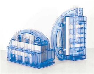
Leukocytes are labeled with BD IMag™ anti-mouse Ly-6G and Ly-6C (Gr-1) Particles - DM according to the following protocol. This labelled cell suspension is then placed within the magnetic field of the BD IMag™ Cell Separation Magnet (Cat. No. 552311). Labelled cells migrate toward the magnet (positive fraction), leaving the unlabelled cells in suspension so they can be drawn off (negative fraction). The tube is then removed from the magnetic field for resuspension of the positive fraction. The separation is repeated twice to increase the purity of the positive fraction. The magnetic separation steps are diagrammed in the Separation Flow Chart. After the positive fraction is washed, the small size of the magnetic particles allows the positive fraction to be further evaluated in downstream applications such as flow cytometry.
MAGNETIC LABELING PROTOCOL
1. Prepare a single-cell suspension from the lymphoid tissue of interest according to standard laboratory procedures. Remove clumps of cells and/or debris by passing the suspension through a 70-µm nylon cell strainer.
2. Dilute BD IMag™ Buffer (10X) (Cat. No. 552362) 1:10 with sterile distilled water to prepare 1X BD IMag buffer by supplementing Phosphate Buffered Saline with 0.5% BSA, 2 mM EDTA, and 0.09% sodium azide). Place on ice.
Although our experience indicates that use of Mouse BD Fc Block™ purified anti-mouse CD16/CD32 mAb 2.4G2 (Cat. No. 553141/553142) is not required for optimal cell separation, some laboratories may want to use it in their studies.
If adding Mouse BD Fc Block, proceed to Step 3.
If not adding Mouse BD Fc Block, proceed to Step 4.
3. Add Mouse BD Fc Block at 0.25 µg/10^6 cells, and incubate on ice for 15 minutes.
4. Wash cells with at least an equal volume of 1X BD IMag buffer, and then resuspend the pellet in 90 µl 1X BD IMag buffer for every 10^7 total cells. If using fewer than 10^7 total cells, resuspend in a 90 µl volume.
5. Vortex the BD IMag™ anti-mouse Ly-6G and Ly-6C (Gr-1) Particles - DM thoroughly, and add 10 µl of particles for every 10^7 total cells.
6. MIX THOROUGHLY. Refrigerate at 6°C - 12°C for 15 minutes.
7. Wash labeled cells with 20 times the labeling volume of 1X BD IMag buffer, remove supernatant completely, and resuspend the cells at a concentration that is appropriate for the magnetic separation column to be used.
8. Separate the cells according to the manufacturer's recommended procedure for the BD IMag™ Cell Separation Magnet being used.
The concentration of BD IMag™ anti-mouse Ly-6G and Ly-6C (Gr-1) Particles - DM suggested in the protocol has been optimized for the purification of Gr-1 positive leukocytes from mouse bone marrow. When labeling target cell populations present at lower frequencies, fewer BD IMag particles can be used. Conversely, when labeling target cell populations that are present at higher frequencies, more particles should be used. To determine the optimal concentration of the BD IMag™ anti-mouse Ly-6G and Ly-6C (Gr-1) Particles - DM for a particular application, a titration in two-fold increments is recommended.
Note: Avoid nonspecific labeling by working quickly and adhering to recommended incubation times.
Product Notices
- Since applications vary, each investigator should titrate the reagent to obtain optimal results.
- Source of all serum proteins is from USDA inspected abattoirs located in the United States.
- Caution: Sodium azide yields highly toxic hydrazoic acid under acidic conditions. Dilute azide compounds in running water before discarding to avoid accumulation of potentially explosive deposits in plumbing.
- BD IMag™ particles are prepared from carboxy-functionalized magnetic particles which are manufactured by Skold Technology and are licensed under US patent number 7,169,618.
- Please refer to www.bdbiosciences.com/us/s/resources for technical protocols.
Data Sheets
Companion Products


BD IMag™ anti-mouse Ly-6G and Ly-6C (gr-1) Particles - DM are magnetic nanoparticles that have monoclonal antibodies conjugated to their surfaces. These particles are optimized for the positive selection or depletion of Gr-1-bearing leukocytes using the BD IMag™ Cell Separation Magnet. In the periphery, RB6-8C5 antibody recognizes primarily granulocytes (neutrophils and eosinophils) and monocytes. In the bone marrow, it recognizes myeloid cells but not erythroid or lymphoid cells.
Development References (4)
-
Conlan JW, North RJ. Neutrophils are essential for early anti-Listeria defense in the liver, but not in the spleen or peritoneal cavity, as revealed by a granulocyte-depleting monoclonal antibody. J Exp Med. 1994; 179(1):259-268. (Biology). View Reference
-
Hestdal K, Ruscetti FW, Ihle JN, et al. Characterization and regulation of RB6-8C5 antigen expression on murine bone marrow cells. J Immunol. 1991; 147(1):22-28. (Biology). View Reference
-
Lagasse E, Weissman IL. Flow cytometric identification of murine neutrophils and monocytes. J Immunol Methods. 1996; 197(1-2):139-150. (Biology). View Reference
-
Tepper RI, Coffman RL, Leder P. An eosinophil-dependent mechanism for the antitumor effect of interleukin-4. Science. 1992; 257(5069):548-551. (Biology). View Reference
Please refer to Support Documents for Quality Certificates
Global - Refer to manufacturer's instructions for use and related User Manuals and Technical data sheets before using this products as described
Comparisons, where applicable, are made against older BD Technology, manual methods or are general performance claims. Comparisons are not made against non-BD technologies, unless otherwise noted.
For Research Use Only. Not for use in diagnostic or therapeutic procedures.
Refer to manufacturer's instructions for use and related User Manuals and Technical Data Sheets before using this product as described.
Comparisons, where applicable, are made against older BD technology, manual methods or are general performance claims. Comparisons are not made against non-BD technologies, unless otherwise noted.