
Preparation And Storage
Recommended Assay Procedures
For optimal and reproducible results, BD Horizon Brilliant™ Stain Buffer should be used anytime BD Horizon Brilliant™ dyes are used in a multicolor flow cytometry panel. Fluorescent dye interactions may cause staining artifacts which may affect data interpretation. The BD Horizon Brilliant Stain Buffer was designed to minimize these interactions. When BD Horizon Brilliant Stain Buffer is used in in the multicolor panel, it should also be used in the corresponding compensation controls for all dyes to achieve the most accurate compensation. For the most accurate compensation, compensation controls created with either cells or beads should be exposed to BD Horizon Brilliant Stain Buffer for the same length of time as the corresponding multicolor panel. More information can be found in the Technical Data Sheet of the BD Horizon Brilliant Stain Buffer (Cat. No. 563794/566349) or the BD Horizon Brilliant Stain Buffer Plus (Cat. No. 566385).
Product Notices
- Since applications vary, each investigator should titrate the reagent to obtain optimal results.
- An isotype control should be used at the same concentration as the antibody of interest.
- Caution: Sodium azide yields highly toxic hydrazoic acid under acidic conditions. Dilute azide compounds in running water before discarding to avoid accumulation of potentially explosive deposits in plumbing.
- Source of all serum proteins is from USDA inspected abattoirs located in the United States.
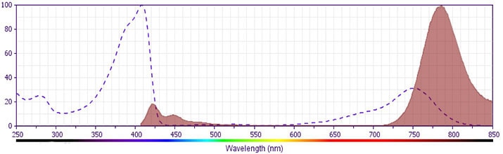
- For fluorochrome spectra and suitable instrument settings, please refer to our Multicolor Flow Cytometry web page at www.bdbiosciences.com/colors.
- BD Horizon Brilliant Violet 786 is covered by one or more of the following US patents: 8,110,673; 8,158,444; 8,227,187; 8,455,613; 8,575,303; 8,354,239.
- BD Horizon Brilliant Stain Buffer is covered by one or more of the following US patents: 8,110,673; 8,158,444; 8,575,303; 8,354,239.
- Cy is a trademark of GE Healthcare.
- Please refer to www.bdbiosciences.com/us/s/resources for technical protocols.
Companion Products


The SB/199 monoclonal antibody specifically binds to mouse CD127 which is also known as, the Interleukin-7 Receptor alpha chain (IL-7Rα). CD127 associates with CD132 (common γ chain) to form a high-affinity signaling IL-7R complex that mediates the biological effects of IL-7. CD127 can also complex with the Thymic Stromal Lymphopoietin Receptor (TSLPR) subunit to bind and mediate cellular responses to TSLP. CD127 is a 65-75 kDa type-I transmembrane protein that belongs to the hematopoietin receptor and the Ig gene superfamilies. Mice lacking CD127 display profoundly impaired development of the B and T lymphoid cell lineages, but display no obvious nonlymphoid abnormalities. IL-7Rα is expressed on common lymphoid progenitors and early stages of B lineage development in the bone marrow, on the earliest thymocyte progenitors, on CD4-CD8- double-negative and CD4+ and CD8+ single-positive thymocytes, and on most peripheral T lymphocytes. Intestinal intraepithelial lymphocytes with low-density γδ TCR expression upregulate CD127 expression in response to IL-2, which may be secreted by neighboring αβ TCR+ T cells.
Development References (9)
-
Akashi K, Kondo M, Weissman IL. Role of interleukin-7 in T-cell development from hematopoietic stem cells. Immunol Rev. 1998; 165:13-28. (Biology). View Reference
-
Faust EA, Saffran DC, Toksoz D, Williams DA, Witte ON. Distinctive growth requirements and gene expression patterns distinguish progenitor B cells from pre-B cells. J Exp Med. 1993; 177(4):915-923. (Biology). View Reference
-
Fujihashi K, Kawabata S, Hiroi T, et al. Interleukin 2 (IL-2) and interleukin 7 (IL-7) reciprocally induce IL-7 and IL-2 receptors on gamma delta T-cell receptor-positive intraepithelial lymphocytes. Proc Natl Acad Sci U S A. 1996; 93(8):3613-3618. (Biology). View Reference
-
Goodwin RG, Friend D, Ziegler SF et al. Cloning of the human and murine interleukin-7 receptors: demonstration of a soluble form and homology to a new receptor superfamily. Cell. 1990; 60(6):941-951. (Biology). View Reference
-
Henderson AJ, Narayanan R, Collins L, Dorshkind K. Status of kappa L chain gene rearrangements and c-kit and IL-7 receptor expression in stromal cell-dependent pre-B cells. J Immunol. 1992; 149(6):1973-1979. (Biology). View Reference
-
Kouro T, Kumar V, Kincade PW. Relationships between early B- and NK-lineage lymphocyte precursors in bone marrow. Blood. 2002; 100(10):3672-3680. (Clone-specific: Flow cytometry, Fluorescence activated cell sorting). View Reference
-
Noguchi M, Nakamura Y, Russell SM, et al. Interleukin-2 receptor gamma chain: a functional component of the interleukin-7 receptor. Science. 1993; 262(5141):1877-1880. (Biology). View Reference
-
Peschon JJ, Morrissey PJ, Grabstein KH, et al. Early lymphocyte expansion is severely impaired in interleukin 7 receptor-deficient mice. J Exp Med. 1994; 180(5):1955-1960. (Biology). View Reference
-
Yamashita Y, Oritani K, Miyoshi EK, Wall R, Bernfield M, Kincade PW. Syndecan-4 is expressed by B lineage lymphocytes and can transmit a signal for formation of dendritic processes. J Immunol. 1999; 162(10):5940-5948. (Clone-specific: Flow cytometry). View Reference
Please refer to Support Documents for Quality Certificates
Global - Refer to manufacturer's instructions for use and related User Manuals and Technical data sheets before using this products as described
Comparisons, where applicable, are made against older BD Technology, manual methods or are general performance claims. Comparisons are not made against non-BD technologies, unless otherwise noted.
For Research Use Only. Not for use in diagnostic or therapeutic procedures.