Skip to main content Skip to navigation
Purified Mouse Anti-NMDAR2B
Western blot analysis of  NMDAR2B on a rat cerebrum lysate (left). Lane 1: 1:250, lane 2: 1:500, lane 3: 1:1000 dilution of the mouse anti- NMDAR2B antibody. Immunofluorescent staining of SK-N-SH cells (right). Cells were seeded in a 384 well collagen coated Microplates (Material # 353962) at ~ 8,000 cells per well.  After overnight incubation, cells were stained using the Triton X100 fix/perm protocol (see Recommended Assay Procedure; Bioimaging protocol link) and the anti- NMDAR2B antibody.  The second step reagent was Alexa Fluor® 488 goat anti mouse Ig (Invitrogen)(pseudo colored green). Cell nuclei were counter stained with Hoechst 33342 (pseudo colored blue). The image was taken on a BD Pathway™  855 or 435 Bioimager System using a 20x objective and merged using the BD AttoVison ™ software. This antibody also stained SH-SY5Y, SK-N-SH, C6, U87 and U373 cells using both the Triton X100 and methanol fix/perm protocols (see Recommended Assay Procedure; Bioimaging protocol link).
Product Details
Down Arrow Up Arrow


BD Transduction Laboratories™
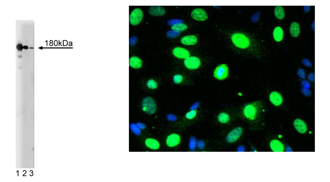
N-Methyl-D-Aspartate Receptor 2B
Rat (QC Testing), Human, Mouse (Tested in Development)
Mouse IgG2b
Rat NMDAR2B aa. 892-1051
Western blot (Routinely Tested), Bioimaging, Immunofluorescence, Immunohistochemistry (Tested During Development), Immunoprecipitation (Not Recommended)
180 kDa
250 µg/ml
AB_397797
Aqueous buffered solution containing BSA, glycerol, and ≤0.09% sodium azide.
RUO


Preparation And Storage

Store undiluted at -20°C. The monoclonal antibody was purified from tissue culture supernatant or ascites by affinity chromatography.

Product Notices

  1. Since applications vary, each investigator should titrate the reagent to obtain optimal results.
  2. Caution: Sodium azide yields highly toxic hydrazoic acid under acidic conditions. Dilute azide compounds in running water before discarding to avoid accumulation of potentially explosive deposits in plumbing.
  3. Source of all serum proteins is from USDA inspected abattoirs located in the United States.
  4. This antibody has been developed and certified for the bioimaging application. However, a routine bioimaging test is not performed on every lot. Researchers are encouraged to titrate the reagent for optimal performance.
  5. Sodium azide is a reversible inhibitor of oxidative metabolism; therefore, antibody preparations containing this preservative agent must not be used in cell cultures nor injected into animals. Sodium azide may be removed by washing stained cells or plate-bound antibody or dialyzing soluble antibody in sodium azide-free buffer. Since endotoxin may also affect the results of functional studies, we recommend the NA/LE (No Azide/Low Endotoxin) antibody format, if available, for in vitro and in vivo use.
  6. Alexa Fluor® is a registered trademark of Molecular Probes, Inc., Eugene, OR.
  7. Triton is a trademark of the Dow Chemical Company.
  8. Please refer to www.bdbiosciences.com/us/s/resources for technical protocols.
610417 Rev. 2
Antibody Details
Down Arrow Up Arrow
13/NMDAR2B

The majority of synapses in the central nervous system utilize glutamate as a neurotransmitter to produce rapid neuronal excitation. Glutamate has a diverse array of receptors that can be categorized into two groups: ionotropic and metabotropic.  The ionotropic receptors are subdivided into two distint types: 1) receptors for N-methyl D-aspartate (NMDAR) and 2) non-NMDA receptors for AMPA and kainate.  Three types of NMDAR2 have been identified:  NR2A, NR2B, and NR2C.  NR2A and NR2B contain a C-terminal extension (>600 amino acids) that has small scattered regions of conserved sequence. The three NR2 mRNAs show overlapping, differential expression patterns in the rat brain.  NR2B has been reported to be expressed in the forebrain, thalamic nuclei, amygdaloid nuclei, caudateputamen, and in restricted regions of the olfactory bulb.

610417 Rev. 2
Format Details
Down Arrow Up Arrow
Purified
Tissue culture supernatant is purified by either protein A/G or affinity purification methods. Both methods yield antibody in solution that is free of most other soluble proteins, lipids, etc. This format provides pure antibody that is suitable for a number of downstream applications including: secondary labeling for flow cytometry or microscopy, ELISA, Western blot, etc.
Purified
610417 Rev.2
Citations & References
Down Arrow Up Arrow

Development References (5)

  1. Fallon L, Moreau F, Croft BG, Labib N, Gu WJ, Fon EA. Parkin and CASK/LIN-2 associate via a PDZ-mediated interaction and are co-localized in lipid rafts and postsynaptic densities in brain. J Biol Chem. 2002; 277(1):486-491. (Biology: Western blot). View Reference
  2. Monyer H, Sprengel R, Schoepfer R. Heteromeric NMDA receptors: molecular and functional distinction of subtypes. Science. 1992; 256(5060):1217-1221. (Biology). View Reference
  3. Sun D, Murali SG. Stimulation of Na+-K+-2Cl- cotransporter in neuronal cells by excitatory neurotransmitter glutamate. Am J Physiol. 1998; 275(3 Pt 1):C772-C779. (Biology: Western blot). View Reference
  4. Wong RW, Setou M, Teng J, Takei Y, Hirokawa N. Overexpression of motor protein KIF17 enhances spatial and working memory in transgenic mice. Proc Natl Acad Sci U S A. 2002; 99(22):14500-14505. (Biology: Western blot). View Reference
  5. Yoshii A, Sheng MH, Constantine-Paton M. Eye opening induces a rapid dendritic localization of PSD-95 in central visual neurons. Proc Natl Acad Sci U S A. 2003; 100(3):1334-1339. (Biology: Immunoprecipitation). View Reference
View All (5) View Less
610417 Rev. 2

Please refer to Support Documents for Quality Certificates


Global - Refer to manufacturer's instructions for use and related User Manuals and Technical data sheets before using this products as described


Comparisons, where applicable, are made against older BD Technology, manual methods or are general performance claims.  Comparisons are not made against non-BD technologies, unless otherwise noted.

For Research Use Only. Not for use in diagnostic or therapeutic procedures.