
-
Your selected country is
Middle East / Africa
- Change country/language
-
Reagents
- Flow Cytometry Reagents
-
Western Blotting and Molecular Reagents
- Immunoassay Reagents
-
Single-Cell Multiomics Reagents
- BD® OMICS-Guard Sample Preservation Buffer
- BD® AbSeq Assay
- BD® OMICS-One Immune Profiler Protein Panel
- BD® Single-Cell Multiplexing Kit
- BD Rhapsody™ ATAC-Seq Assays
- BD Rhapsody™ Whole Transcriptome Analysis (WTA) Amplification Kit
- BD Rhapsody™ TCR/BCR Next Multiomic Assays
- BD Rhapsody™ Targeted mRNA Kits
- BD Rhapsody™ Accessory Kits
-
Functional Assays
-
Microscopy and Imaging Reagents
-
Cell Preparation and Separation Reagents
-
- BD® OMICS-Guard Sample Preservation Buffer
- BD® AbSeq Assay
- BD® OMICS-One Immune Profiler Protein Panel
- BD® Single-Cell Multiplexing Kit
- BD Rhapsody™ ATAC-Seq Assays
- BD Rhapsody™ Whole Transcriptome Analysis (WTA) Amplification Kit
- BD Rhapsody™ TCR/BCR Next Multiomic Assays
- BD Rhapsody™ Targeted mRNA Kits
- BD Rhapsody™ Accessory Kits
- Middle East / Africa (English)
-
Change country/language
Old Browser
This page has been recently translated and is available in French now.
Looks like you're visiting us from {countryName}.
Would you like to stay on the current country site or be switched to your country?


Western blot and flow cytometric analyses of ROR2 expression in human chronic myelogenous leukemia cell line. LEFT: Lysate prepared from K562 cells (ATCC CCL-243) was electrophoresed (SDS-PAGE), transferred to PVDF membranes and probed with 500, 250, 125 and 62.5 ng/mL (lanes 1-4, respectively) of Purified Mouse Anti-ROR2 (Cat. No. 565550). HRP Goat Anti-Mouse Ig (Cat. No. 554002) was used to develop the specific staining. Human ROR2 was identified as a band whose size ranged from 100-150 kDa. RIGHT: K562 cells (ATCC CCL-243) were fixed in BD Cytofix™ fixation buffer (Cat. No. 554655) and permeabilized in BD Perm/Wash™ buffer (Cat. No. 554723). Permeablized cells were stained with either Purified Mouse IgG1 κ Isotype Control (Cat. No. 554121, dashed line) or Purified Mouse Anti-ROR2 (Cat. No. 565550, solid line), followed by PE Goat Anti-Mouse Ig (Cat. No. 550589). The fluorescence histograms were derived from gated events with the forward and side light-scatter characteristics of viable cells. Flow cytometry was performed using a BD FACSCanto™ II Cell Analyzer.

Immunohistochemical staining of ROR2 in human and rat tissue sections. Following antigen retrieval with BD Retrievagen A Buffer (Cat. No. 550524), the formalin-fixed paraffin-embedded sections were stained with either Purified Mouse IgG1 κ Isotype Control (Cat. No. 550878; left column) or Purified Mouse Anti-ROR2 antibody (Cat. No. 565550; right column). A three-step staining procedure that employs a Biotin Goat Anti-Mouse Immunoglobulin (Cat. No. 550337), Streptavidin-Horseradish Peroxidase (HRP) (Cat. No.550946), and the DAB Substrate Kit (Cat. No. 550880) was used to develop the primary staining reagents. Top Row: Human cerebellum. Original magnification: 10× Middle Row: Rat cerebellum. Original magnification: 20× Bottom Row: Rat embryo. Original magnification: 20×


BD Pharmingen™ Purified Mouse Anti-ROR2

BD Pharmingen™ Purified Mouse Anti-ROR2

Regulatory Status Legend
Any use of products other than the permitted use without the express written authorization of Becton, Dickinson and Company is strictly prohibited.
Preparation And Storage
Product Notices
- Since applications vary, each investigator should titrate the reagent to obtain optimal results.
- Please refer to www.bdbiosciences.com/us/s/resources for technical protocols.
- Caution: Sodium azide yields highly toxic hydrazoic acid under acidic conditions. Dilute azide compounds in running water before discarding to avoid accumulation of potentially explosive deposits in plumbing.
- Sodium azide is a reversible inhibitor of oxidative metabolism; therefore, antibody preparations containing this preservative agent must not be used in cell cultures nor injected into animals. Sodium azide may be removed by washing stained cells or plate-bound antibody or dialyzing soluble antibody in sodium azide-free buffer. Since endotoxin may also affect the results of functional studies, we recommend the NA/LE (No Azide/Low Endotoxin) antibody format, if available, for in vitro and in vivo use.
- An isotype control should be used at the same concentration as the antibody of interest.
- Species cross-reactivity detected in product development may not have been confirmed on every format and/or application.
Companion Products


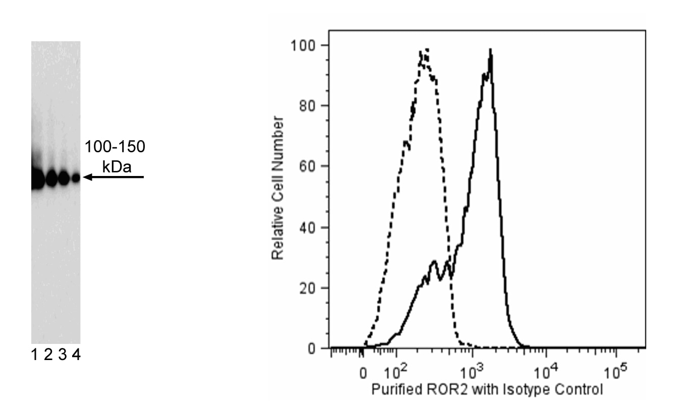
The Ror2 monoclonal antibody specifically binds to the ST2 intracellular domain of ROR2 (Receptor tyrosine kinase-like Orphan Receptor 2). ROR2 is a type I transmembrane protein that belongs to a small family of receptor tyrosine kinases containing frizzled domains. Based on the homology of ROR2 and Wnt receptors Frz domains, ROR2 has been linked to Wnt signaling and embryonic development. Wnt5a-ROR2 signaling promotes osteoclastogenesis, and ROR2 is also critical for the formation of skeleton, heart, and genitals. In normal adult tissues, ROR2 is expressed in very low levels or undetectable. There is mounting evidence that ROR2 expression is associated with either tumor progression or suppression in different tissues. In humans, mutations in the ROR2 gene are associated with the autosomal recessive form of Robinow Syndrome and autosomal dominant Brachydactyly type B.
Development References (3)
-
Ford CE, Qian Ma SS, Quadir A, Ward RL. The dual role of the novel Wnt receptor tyrosine kinase, ROR2, in human carcinogenesis. Int J Cancer. 2013; 133(4):779-787. (Biology). View Reference
-
Mikels A, Minami Y, Nusse R. Ror2 receptor requires tyrosine kinase activity to mediate Wnt5A signaling. J Biol Chem. 2009; 284(44):30167-30176. (Immunogen: Immunohistochemistry, Western blot). View Reference
-
van Amerongen R, Fuerer C, Mizutani M, Nusse R. Wnt5a can both activate and repress Wnt/β-catenin signaling during mouse embryonic development. Dev Biol. 2012; 369(1):101-114. (Clone-specific: Immunohistochemistry, Western blot). View Reference
Please refer to Support Documents for Quality Certificates
Global - Refer to manufacturer's instructions for use and related User Manuals and Technical data sheets before using this products as described
Comparisons, where applicable, are made against older BD Technology, manual methods or are general performance claims. Comparisons are not made against non-BD technologies, unless otherwise noted.
For Research Use Only. Not for use in diagnostic or therapeutic procedures.