
Preparation And Storage
Product Notices
- This reagent has been pre-diluted for use at the recommended Volume per Test. We typically use 1 × 10^6 cells in a 100-µl experimental sample (a test).
- An isotype control should be used at the same concentration as the antibody of interest.
- Caution: Sodium azide yields highly toxic hydrazoic acid under acidic conditions. Dilute azide compounds in running water before discarding to avoid accumulation of potentially explosive deposits in plumbing.
- Source of all serum proteins is from USDA inspected abattoirs located in the United States.
- Please observe the following precautions: Absorption of visible light can significantly alter the energy transfer occurring in any tandem fluorochrome conjugate; therefore, we recommend that special precautions be taken (such as wrapping vials, tubes, or racks in aluminum foil) to prevent exposure of conjugated reagents, including cells stained with those reagents, to room illumination.
- PerCP-Cy5.5–labelled antibodies can be used with FITC- and R-PE–labelled reagents in single-laser flow cytometers with no significant spectral overlap of PerCP-Cy5.5, FITC, and R-PE fluorescence.
- PerCP-Cy5.5 is optimized for use with a single argon ion laser emitting 488-nm light. Because of the broad absorption spectrum of the tandem fluorochrome, extra care must be taken when using dual-laser cytometers, which may directly excite both PerCP and Cy5.5™. We recommend the use of cross-beam compensation during data acquisition or software compensation during data analysis.
- For fluorochrome spectra and suitable instrument settings, please refer to our Multicolor Flow Cytometry web page at www.bdbiosciences.com/colors.
- Cy is a trademark of GE Healthcare.
- Please refer to http://regdocs.bd.com to access safety data sheets (SDS).
- Please refer to www.bdbiosciences.com/us/s/resources for technical protocols.
Companion Products


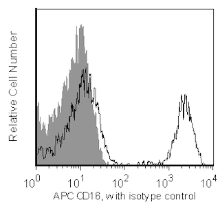
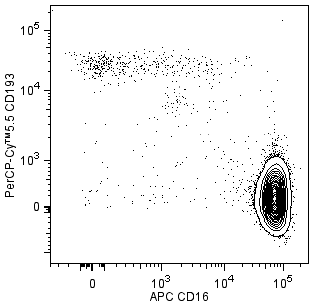
The 5E8 monoclonal antibody specifically binds to human CCR3 which is also known as CD193. CCR3 is a G protein-linked, 7 transmembrane, chemokine receptor expressed on a variety of hematopoietic cells. Similar to CCR5 and CXCR4, CCR3 can be a co-receptor for HIV-1. It is primarily expressed by eosinophils and basophils during atopic conditions, dermatitis, allergic rhinitis, conjunctivitis and bronchial asthma. Chemokines including RANTES, Eotaxin, MCP-3, MIP1α have been reported to act as ligands for CCR3 and stimulate CCR3+ cells. Eotaxin stimulates Th2 cells expressing CCR3. Other studies describe HIV-1 specific T cell cytotoxicity can be mediated by RANTES and Eotaxin through CCR3. CCR3 expressed on dendritic cells may have a biological role on cell-cell interaction during antigen presentation. CCR3 has been clustered as CD193 in the HLDA VIIIth workshop.

Development References (9)
-
Agrawal L, Maxwell CR, Peters PJ, et al. Complexity in human immunodeficiency virus type 1 (HIV-1) co-receptor usage: roles of CCR3 and CCR5 in HIV-1 infection of monocyte-derived macrophages and brain microglia. J Gen Virol. 2009; 90(3):710-722. (Clone-specific: Flow cytometry, Immunofluorescence). View Reference
-
Daugherty BL, Siciliano SJ, DeMartino JA, Malkowitz L, Sirotina A, Springer MS. Cloning, expression, and characterization of the human eosinophil eotaxin receptor. J Exp Med. 1996; 83(5):2349-2354. (Biology). View Reference
-
Ghorpade A, Xia MQ, Hyman BT, et al. Role of the beta-chemokine receptors CCR3 and CCR5 in human immunodeficiency virus type 1 infection of monocytes and microglia. J Virol. 1998; 72(4):3351-3361. (Biology). View Reference
-
Hadida F, Vieillard V, Autran B, Clark-Lewis I, Baggiolini M, Debre P. HIV-specific T cell cytotoxicity mediated by RANTES via the chemokine receptor CCR3. J Exp Med. 1998; 188(3):609-614. (Biology). View Reference
-
Heath H, Qin S, Rao P, et al. Chemokine receptor usage by human eosinophils. The importance of CCR3 demonstrated using an antagonistic monoclonal antibody. J Clin Invest. 1997; 99(2):178-184. (Immunogen: Flow cytometry). View Reference
-
Liu SM, Xavier R, Good KL, et al. Immune cell transcriptome datasets reveal novel leukocyte subset-specific genes and genes associated with allergic processes. J Allergy Clin Immunol. 2006; 118(2):496-503. (Clone-specific). View Reference
-
Sallusto F, Mackay CR, Lanzavecchia A. Selective expression of the eotaxin receptor CCR3 by human T helper 2 cells. Science. 1997; 277(5334):2005-2007. (Biology). View Reference
-
Sato K, Kawasaki H, Nagayama H, et al. CC chemokine receptors, CCR-1 and CCR-3, are potentially involved in antigen-presenting cell function of human peripheral blood monocyte-derived dendritic cells. Blood. 1999; 93(1):34-42. (Biology). View Reference
-
Zimmermann N, Daugherty BL, Stark JM, Rothenberg ME. Molecular analysis of CCR-3 events in eosinophilic cells. J Immunol. 2000; 164(2):1055-1064. (Biology). View Reference
Please refer to Support Documents for Quality Certificates
Global - Refer to manufacturer's instructions for use and related User Manuals and Technical data sheets before using this products as described
Comparisons, where applicable, are made against older BD Technology, manual methods or are general performance claims. Comparisons are not made against non-BD technologies, unless otherwise noted.
For Research Use Only. Not for use in diagnostic or therapeutic procedures.