-
Your selected country is
Middle East / Africa
- Change country/language
Old Browser
This page has been recently translated and is available in French now.
Looks like you're visiting us from {countryName}.
Would you like to stay on the current country site or be switched to your country?


.png)

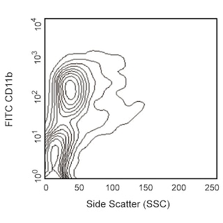
Multicolor flow cytometric analysis of Notch1 expression on BALB/c mouse bone marrow cells and thymocytes. Left Panel: Mouse bone marrow cells were stained with FITC Rat Anti-Mouse CD11b antibody (Cat. No. 553310/557396/561688) and either PE Rat IgG2a, κ Isotype Control (Cat. No. 554689, Left Plot) or PE Rat Anti-Mouse Notch1 antibody (Cat. No. 562754, Right Plot). The two-color flow cytometric dot plots showing the expression of CD11b versus Notch1 (or Ig Isotype Control staining) were derived from gated events with the forward and side light-scatter characteristics of viable bone marrow cells. Right Panel: Thymocytes were stained with PE Rat Anti-Mouse Notch1, PE-Cy™7 Rat Anti-Mouse CD4 (Cat. No. 552775) and APC Rat Anti-Mouse CD8a (Cat. No. 553035) antibodies. The dot plot shows the expression of CD8 versus CD4 by viable thymocytes with Notch1-positive thymocytes colorized red. Flow cytometry was performed using a BD FACSCanto™ II System.
.png)

BD Pharmingen™ PE Rat anti-Mouse Notch1
.png)
Regulatory Status Legend
Any use of products other than the permitted use without the express written authorization of Becton, Dickinson and Company is strictly prohibited.
Preparation And Storage
Product Notices
- Since applications vary, each investigator should titrate the reagent to obtain optimal results.
- An isotype control should be used at the same concentration as the antibody of interest.
- Caution: Sodium azide yields highly toxic hydrazoic acid under acidic conditions. Dilute azide compounds in running water before discarding to avoid accumulation of potentially explosive deposits in plumbing.
- For fluorochrome spectra and suitable instrument settings, please refer to our Multicolor Flow Cytometry web page at www.bdbiosciences.com/colors.
- Cy is a trademark of GE Healthcare.
- Please refer to www.bdbiosciences.com/us/s/resources for technical protocols.
Companion Products

.png?imwidth=320)


.png?imwidth=320)
The 22E5.5 monoclonal antibody specifically binds to the extracellular domain of mouse Notch1. This Type 1 transmembrane protein is a member of the Notch family that includes Notch1-4. Notch ligands include membrane-bound Jagged1, Jagged2 and Delta-like1, 3 and 4. Notch receptors mediate juxtacrine signals between adjacent cells. Upon ligand binding, Notch1 undergoes proteolytic cleavage that results in the release of the Notch intracellular domain, NICD. NICD translocates to the nucleus where it forms a transcriptional activator complex with the RBP-J DNA binding factor and other transcriptional cofactors. These multimeric complexes regulate the expression of multiple genes including those that orchestrate many facets of embryonic development and the subsequent functioning of multiple organ systems such as the immune and nervous systems. Within the immune system, Notch signaling significantly affects the differentiation and survival of numerous cell types including thymocytes and subsets of T and B lymphocytes and dendritic cells. In altered forms, Notch1 has been implicated in transformation or progression in some T-cell neoplasms. The mN1A monoclonal antibody specifically binds to an intracellular domain of mouse Notch1.

Development References (7)
-
Anderson AC, Robey EA, Huang YH. Notch signaling in lymphocyte development. Curr Opin Genet Dev. 2001; 11(5):554-560. (Biology). View Reference
-
Artavanis-Tsakonas S, Rand MD, Lake RJ. Notch signaling: cell fate control and signal integration in development.. Science. 1999; 284(5415):770-6. (Biology). View Reference
-
Fiorini E, Merck E, Wilson A, et al. Dynamic regulation of Notch1 and Notch2 surface expression during T cell development and activation revealed by novel monoclonal antibodies. J Immunol. 2009; 183(11):7212-7222. (Immunogen: Flow cytometry, Immunofluorescence). View Reference
-
Helbig C, Gentek R, Backer RA, et al. Notch controls the magnitude of T helper cell responses by promoting cellular longevity. Proc Natl Acad Sci U S A. 109(23):9041-9046. (Biology). View Reference
-
Kojika S, Griffin JD. Notch receptors and hematopoiesis. Exp Hematol. 2001; 29(9):1041-1052. (Biology). View Reference
-
Radtke F, Fasnacht N, Macdonald HR. Notch signaling in the immune system. Immunity. 2010; 32(1):14-27. (Biology). View Reference
-
Walker L, Carlson A, Tan-Pertel HT, Weinmaster G, Gasson J. The notch receptor and its ligands are selectively expressed during hematopoietic development in the mouse. Stem Cells. 2001; 19(6):543-552. (Biology). View Reference
Please refer to Support Documents for Quality Certificates
Global - Refer to manufacturer's instructions for use and related User Manuals and Technical data sheets before using this products as described
Comparisons, where applicable, are made against older BD Technology, manual methods or are general performance claims. Comparisons are not made against non-BD technologies, unless otherwise noted.
For Research Use Only. Not for use in diagnostic or therapeutic procedures.
Report a Site Issue
This form is intended to help us improve our website experience. For other support, please visit our Contact Us page.