Skip to main content Skip to navigation
PE Rat Anti-Mouse IL-4
Expression of IL-4 by stimulated CD4+ and CD4-BALB/c spleen cells.  Splenocytes from 6 month old BALB/c mice were cultured for 72 hours in medium containing Staphylococcus aureus enterotoxin B (2 µg/mL final concentration; Sigma, St. Louis, MO), recombinant mouse IL-2 (10 U/mL final concentration; Cat. No. 550069) and recombinant mouse IL-4 (2 ng/mL final concentration; Cat. No. 550067). The cells were harvested and restimulated for 5 hours with anti-CD3 (2 µg/mL final concentration; 145-2C11, Cat. No. 553057) and anti-CD28 (2 µg/mL final concentration; clone 37.51, Cat. No. 553294) antibodies in the presence of GolgiStop™ (2 µM final concentration; (Cat. No. 554724). The splenocytes were harvested, stained with 0.06 µg of FITC-conjugated rat anti-mouse CD4 (FITC-RM4-5, Cat. No. 553047) fixed, permeabilized, and subsequently stained with 0.25 µg of PE-conjugated rat anti-mouse IL-4 antibody (PE-BVD4-1D11, Cat. No. 554389) by using the BD Pharmingen staining protocol (see left panel). To demonstrate specificity of staining, the binding of PE-BVD4-1D11 was blocked by the preincubation of the conjugated antibody with recombinant mouse IL-4 (0.25 µg, Cat. No. 550067; see middle panel), and by preincubation of the fixed/permeabilized cells with unlabelled BVD4-1D11 antibody (5 µg, Cat. No. 554386; see right panel). The quadrant markers for the bivariate dot plots were set based on the autofluorescence control, and verified with the recombinant cytokine blocking and unlabelled antibody blocking specificity controls.
Product Details
Down Arrow Up Arrow


BD Pharmingen™
Mouse (QC Testing)
Rat IgG2b
Recombinant Mouse IL-4
Intracellular staining (flow cytometry) (Routinely Tested)
0.2 mg/ml
AB_395361
Aqueous buffered solution containing ≤0.09% sodium azide.
RUO


Preparation And Storage

Store undiluted at 4°C and protected from prolonged exposure to light. Do not freeze. The monoclonal antibody was purified from tissue culture supernatant or ascites by affinity chromatography. The antibody was conjugated with R-PE under optimum conditions, and unconjugated antibody and free PE were removed.

Product Notices

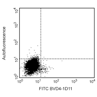
  1. Since applications vary, each investigator should titrate the reagent to obtain optimal results.
  2. Caution: Sodium azide yields highly toxic hydrazoic acid under acidic conditions. Dilute azide compounds in running water before discarding to avoid accumulation of potentially explosive deposits in plumbing.
  3. For fluorochrome spectra and suitable instrument settings, please refer to our Multicolor Flow Cytometry web page at www.bdbiosciences.com/colors.
  4. Please refer to www.bdbiosciences.com/us/s/resources for technical protocols.
554389 Rev. 3
Antibody Details
Down Arrow Up Arrow
BVD4-1D11

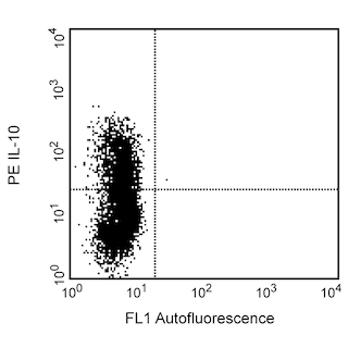
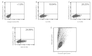
IL-4 is a pleiotropic cytokine that can regulate immunological responses including cell proliferation, survival and gene expression. Expressed by mast cells, basophils, eosinophils and T-cells, IL-4 regulates the differentiation of naive CD4+ T cells into helper Th2 cells.  IL-4 can also be involved with the regulation of immunoglobulin class switching to the IgG1 and IgE isotypes in addition to being involved with the development of allergic inflammation and asthma.

554389 Rev. 3
Format Details
Down Arrow Up Arrow
PE
R-Phycoerythrin (PE), is part of the BD family of Phycobiliprotein dyes. This fluorochrome is a multimeric fluorescent phycobiliprotein with excitation maximum (Ex Max) of 496 nm and 566 nm and an emission maximum (Em Max) at 576 nm. PE is designed to be excited by the Blue (488 nm), Green (532 nm) and Yellow-Green (561 nm) lasers and detected using an optical filter centered near 575 nm (e.g., a 575/26-nm bandpass filter). As PE is excited by multiple lasers, this can result in cross-laser excitation and fluorescence spillover on instruments with various combinations of Blue, Green, and Yellow-Green lasers. Please ensure that your instrument’s configurations (lasers and optical filters) are appropriate for this dye.
altImg
PE
Yellow-Green 488 nm, 532 nm, 561 nm
496 nm, 566 nm
576 nm
554389 Rev.3
Citations & References
Down Arrow Up Arrow

Development References (5)

  1. Abrams JS, Roncarolo MG, Yssel H, Andersson U, Gleich GJ, Silver JE. Strategies of anti-cytokine monoclonal antibody development: immunoassay of IL-10 and IL-5 in clinical samples. Immunol Rev. 1992; 127:5-24. (Clone-specific: ELISA, Neutralization). View Reference
  2. Finkelman FD, Madden KB, Morris SC, et al. Anti-cytokine antibodies as carrier proteins. Prolongation of in vivo effects of exogenous cytokines by injection of cytokine-anti-cytokine antibody complexes. J Immunol. 1993; 151(3):1235-1244. (Clone-specific: ELISA, Neutralization). View Reference
  3. Litton MJ, Sander B, Murphy E, O'Garra A, Abrams JS. Early expression of cytokines in lymph nodes after treatment in vivo with Staphylococcus enterotoxin B. J Immunol Methods. 1994; 175(1):47-58. (Clone-specific: ELISA). View Reference
  4. Prussin C, Metcalfe DD. Detection of intracytoplasmic cytokine using flow cytometry and directly conjugated anti-cytokine antibodies. J Immunol Methods. 1995; 188(1):117-128. (Biology). View Reference
  5. Sander B, Hoiden I, Andersson U, Moller E, Abrams JS. Similar frequencies and kinetics of cytokine producing cells in murine peripheral blood and spleen. Cytokine detection by immunoassay and intracellular immunostaining. J Immunol Methods. 1993; 166(2):201-214. (Clone-specific: ELISA, Neutralization). View Reference
View All (5) View Less
554389 Rev. 3

Please refer to Support Documents for Quality Certificates


Global - Refer to manufacturer's instructions for use and related User Manuals and Technical data sheets before using this products as described


Comparisons, where applicable, are made against older BD Technology, manual methods or are general performance claims.  Comparisons are not made against non-BD technologies, unless otherwise noted.

For Research Use Only. Not for use in diagnostic or therapeutic procedures.