Skip to main content Skip to navigation
BV421 Rat Anti-Mouse CD279 (PD-1)
Alert icon
This product is the replacement for 748268.
BV421 Rat Anti-Mouse CD279 (PD-1)
Flow cytometric analysis of CD279 (PD-1) expression on resting and activated Mouse splenic leukocytes.  C57BL/6 Mouse splenocytes were cultured with plate-bound Purified NA/LE Hamster Anti-Mouse CD3e antibody (Cat. No. 553057) for 3 days. The resting (Left Plot) or activated (Right Plot) splenocytes were harvested, preincubated with Purified Rat Anti-Mouse CD16/CD32 antibody (Mouse BD Fc Block™) [Cat. No. 553141/553142], and then stained with either BD Horizon™ BV421 Rat IgG2b, κ Isotype Control (Cat. No. 562603; dashed line histograms) or BD Horizon™ BV421 Rat Anti-Mouse CD279 (PD-1) antibody (Cat. No. 569780; solid line histograms) at 0.5 μg/test. BD Via-Probe™ Cell Viability 7-AAD Solution (Cat. No. 555815/555816) was added to cells right before analysis. The fluorescence histograms showing CD279 (PD-1) expression (or Ig Isotype control staining) were derived from gated events with the forward and side light-scatter characteristics of viable (7-AAD-negative) splenic leukocytes. Flow cytometry and data analysis were performed using a BD LSRFortessa™ X-20 Cell Analyzer System and FlowJo™ software. Data shown on this Technical Data Sheet are not lot specific.
Product Details
Down Arrow Up Arrow


BD Horizon™
PD-1; mPD-1; Pdc1; Pdcd1; Ly101
Mouse (QC Testing)
Rat SD, also known as Sprague-Dawley (outbred) IgG2b, κ
Mouse PD-1 Recombinant Protein
Flow cytometry (Routinely Tested)
0.2 mg/ml
18566
Aqueous buffered solution containing ≤0.09% sodium azide.
RUO


Preparation And Storage

The monoclonal antibody was purified from tissue culture supernatant or ascites by affinity chromatography. The antibody was conjugated to the dye under optimum conditions and unconjugated antibody and free dye were removed. Store undiluted at 4°C and protected from prolonged exposure to light. Do not freeze.

Recommended Assay Procedures

   BD® CompBeads can be used as surrogates to assess fluorescence spillover (compensation).  When fluorochrome conjugated antibodies are bound to BD® CompBeads, they have spectral properties very similar to cells.   However, for some fluorochromes there can be small differences in spectral emissions compared to cells, resulting in spillover values that differ when compared to biological controls.  It is strongly recommended that when using a reagent for the first time, users compare the spillover on cells and BD® CompBeads to ensure that BD® CompBeads are appropriate for your specific cellular application.

   For optimal and reproducible results, BD Horizon Brilliant Stain Buffer should be used anytime BD Horizon Brilliant dyes are used in a multicolor flow cytometry panel.  Fluorescent dye interactions may cause staining artifacts which may affect data interpretation.  The BD Horizon Brilliant Stain Buffer was designed to minimize these interactions. When BD Horizon Brilliant Stain Buffer is used in the multicolor panel, it should also be used in the corresponding compensation controls for all dyes to achieve the most accurate compensation. For the most accurate compensation, compensation controls created with either cells or beads should be exposed to BD Horizon Brilliant Stain Buffer for the same length of time as the corresponding multicolor panel. More information can be found in the Technical Data Sheet of the BD Horizon Brilliant Stain Buffer (Cat. No. 563794/566349) or the BD Horizon Brilliant Stain Buffer Plus (Cat. No. 566385).

Product Notices

  1. Please refer to www.bdbiosciences.com/us/s/resources for technical protocols.
  2. Since applications vary, each investigator should titrate the reagent to obtain optimal results.
  3. An isotype control should be used at the same concentration as the antibody of interest.
  4. Caution: Sodium azide yields highly toxic hydrazoic acid under acidic conditions. Dilute azide compounds in running water before discarding to avoid accumulation of potentially explosive deposits in plumbing.
  5. For fluorochrome spectra and suitable instrument settings, please refer to our Multicolor Flow Cytometry web page at www.bdbiosciences.com/colors.
  6. BD Horizon Brilliant Violet 421 is covered by one or more of the following US patents: 8,158,444; 8,362,193; 8,575,303; 8,354,239.
  7. BD Horizon Brilliant Stain Buffer is covered by one or more of the following US patents: 8,110,673; 8,158,444; 8,575,303; 8,354,239.
  8. Please refer to http://regdocs.bd.com to access safety data sheets (SDS).
  9. For U.S. patents that may apply, see bd.com/patents.
569780 Rev. 1
Antibody Details
Down Arrow Up Arrow
RMP1-30

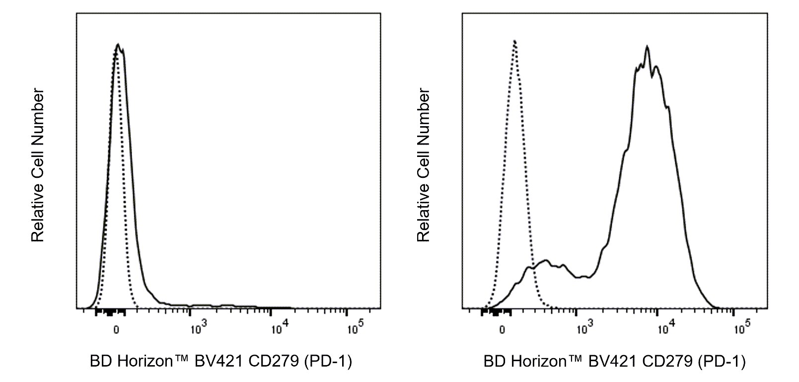
The RMP1-30 monoclonal antibody specifically recognizes CD279 which is also known as PD-1 (programmed death-1). CD279 (PD-1) is a ~55 kDa type I transmembrane glycoprotein that is encoded by Pdcd1 which belongs to the CD28/CTLA-4 family within the Ig superfamily. CD279 (PD-1) is comprised of an extracellular region with an IgV-like domain and an intracellular region with an immunoreceptor tyrosine-based inhibitory motif (ITIM) and an immunoreceptor tyrosine-based switch motif (ITSM) that are associated with inhibitory signaling functions. CD279 (PD-1) is transiently expressed on CD4-CD8- thymocytes and developing B lymphocytes at the pro-B-cell stage. It is also expressed on activated myeloid cells, B cells, and T cells including exhausted T cells found in mice during chronic viral infections or cancer. This co-inhibitory receptor reportedly functions in negative regulation of immune responses and thus helps guard against autoimmunity and preserves peripheral tolerance. CD273 (also known as PD-L2 or B7-DC) and CD274 (PD-L1 or B7-H1) are members of the B7 family within the Ig superfamily that serve as ligands for CD279 (PD-1). The RMP1-30 antibody reportedly does not block the binding of CD279 (PD-1) to these ligands.

569780 Rev. 1
Format Details
Down Arrow Up Arrow
BV421
The BD Horizon Brilliant Violet™ 421 (BV421) Dye is part of the BD Horizon Brilliant Violet™ family of dyes. This polymer-technology based dye has an excitation maximum (Ex Max) of 407-nm and an emission maximum (Em Max) at 423-nm. Driven by BD innovation, BV421 is designed to be excited by the violet laser (405-nm) and detected using an optical filter centered near 420-nm (e.g., a 431/28-nm or 450/50-nm bandpass filter). BV421 is an ideal alternative for V450 as it is approximately ten times brighter with less spillover into the BV510/V500 detector. Please ensure that your instrument’s configurations (lasers and optical filters) are appropriate for this dye.
altImg
BV421
Violet 405 nm
407 nm
423 nm
569780 Rev.1
Citations & References
Down Arrow Up Arrow

Development References (7)

  1. Agata Y, Kawasaki A, Nishimura H, et al. Expression of the PD-1 antigen on the surface of stimulated mouse T and B lymphocytes. Int Immunol. 1996 May; 8(5):765-772. (Biology). View Reference
  2. Gupta PK, Godec J, Wolski D, et al. CD39 Expression Identifies Terminally Exhausted CD8+ T Cells.. PLoS Pathog. 2015; 11(10):e1005177. (Clone-specific: Flow cytometry). View Reference
  3. Kasagi S, Kawano S, Okazaki T, et al. Anti-programmed cell death 1 antibody reduces CD4+PD-1+ T cells and relieves the lupus-like nephritis of NZB/W F1 mice.. J Immunol. 2010; 184(5):2337-47. (Clone-specific: Flow cytometry). View Reference
  4. Liu X, Gibbons RM, Harrington SM, et al. Endogenous tumor-reactive CD8+ T cells are differentiated effector cells expressing high levels of CD11a and PD-1 but are unable to control tumor growth.. Oncoimmunology. 2013; 2(6):e23972. (Clone-specific: Flow cytometry). View Reference
  5. Matsumoto K, Inoue H, Nakano T, et al. B7-DC regulates asthmatic response by an IFN-gamma-dependent mechanism.. J Immunol. 2004; 172(4):2530-41. (Immunogen). View Reference
  6. Nishimura H, Agata Y, Kawasaki A, et al. Developmentally regulated expression of the PD-1 protein on the surface of double-negative (CD4-CD8-) thymocytes. Int Immunol. 1996 May; 8(5):773-780. (Biology). View Reference
  7. Tsushima F, Iwai H, Otsuki N, et al. Preferential contribution of B7-H1 to programmed death-1-mediated regulation of hapten-specific allergic inflammatory responses. Eur J Immunol. 2003; 33(10):2773-2782. (Biology). View Reference
View All (7) View Less
569780 Rev. 1

Please refer to Support Documents for Quality Certificates


Global - Refer to manufacturer's instructions for use and related User Manuals and Technical data sheets before using this products as described


Comparisons, where applicable, are made against older BD Technology, manual methods or are general performance claims.  Comparisons are not made against non-BD technologies, unless otherwise noted.

For Research Use Only. Not for use in diagnostic or therapeutic procedures.