







Preparation And Storage


Recommended Assay Procedures
For optimal and reproducible results, BD Horizon Brilliant Stain Buffer should be used anytime two or more BD Horizon Brilliant dyes are used in the same experiment. Fluorescent dye interactions may cause staining artifacts which may affect data interpretation. The BD Horizon Brilliant Stain Buffer was designed to minimize these interactions. More information can be found in the Technical Data Sheet of the BD Horizon Brilliant Stain Buffer (Cat. No. 563794/566349).
Product Notices
- This reagent has been pre-diluted for use at the recommended Volume per Test. We typically use 1 × 10^6 cells in a 100-µl experimental sample (a test).
- An isotype control should be used at the same concentration as the antibody of interest.
- Source of all serum proteins is from USDA inspected abattoirs located in the United States.
- Caution: Sodium azide yields highly toxic hydrazoic acid under acidic conditions. Dilute azide compounds in running water before discarding to avoid accumulation of potentially explosive deposits in plumbing.
- Pacific Blue™ is a trademark of Molecular Probes, Inc., Eugene, OR.
- Accutase is a registered trademark of Innovative Cell Technologies, Inc.
- For fluorochrome spectra and suitable instrument settings, please refer to our Multicolor Flow Cytometry web page at www.bdbiosciences.com/colors.
- BD Horizon Brilliant Violet 421 is covered by one or more of the following US patents: 8,158,444; 8,362,193; 8,575,303; 8,354,239.
- BD Horizon Brilliant Stain Buffer is covered by one or more of the following US patents: 8,110,673; 8,158,444; 8,575,303; 8,354,239.
- Please refer to www.bdbiosciences.com/us/s/resources for technical protocols.
Companion Products




Gangliosides are sialic-acid bearing glycolipids that are expressed on the surface of all mammalian cells, and are likely involved in mediating cell-substratum interactions. They are important target antigens for antibody dependent cellular cytotoxicity (ADCC) of human melanoma and neuroblastoma cells. Human melanoma cells produce gangliosides, designated as GD2 and GD3 which are deposited in the subtratum-attached material, and may play a significant role in the melanoma metastatic phenotype. Clone 14.G2a specifically reacts with human and mouse GD2 ganglioside. LAN-1 human neuroblastoma cells were used as immunogen. Clone 14.G2a is an isotype switch variant selected from the parental IgG3-producing hybridoma 14.18 and has identical reactivity as the parental antibody. Clone 14.G2a is routinely tested by flow cytometry using M21 human melanoma cells.
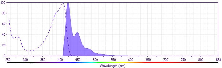
The antibody was conjugated to BD Horizon™ BV421 which is part of the BD Horizon Brilliant™ Violet family of dyes. With an Ex Max of 407-nm and Em Max at 421-nm, BD Horizon BV421 can be excited by the violet laser and detected in the standard Pacific Blue™ filter set (eg, 450/50-nm filter). BD Horizon BV421 conjugates are very bright, often exhibiting a 10 fold improvement in brightness compared to Pacific Blue conjugates.
Development References (6)
-
Cheresh DA, Klier FG. Disialoganglioside GD2 distributes preferentially into substrate-associated microprocesses on human melanoma cells during their attachment to fibronectin. J Cell Biol. 1986; 102(5):1887-1897. (Biology). View Reference
-
Frost JD, Hank JA, Reaman GH, et al. A phase I/IB trial of murine monoclonal anti-GD2 antibody 14.G2a plus interleukin-2 in children with refractory neuroblastoma: a report of the Children's Cancer Group. Cancer. 1997; 80(2):317-333. (Clone-specific: Cytotoxicity, In vivo exacerbation). View Reference
-
Hakomori S. Tumor-associated carbohydrate antigens. Annu Rev Immunol. 1984; 2:103-126. (Biology). View Reference
-
Lode HN, Reisfeld RA, Handgretinger R, Nicolaou KC, Gaedicke G, Wrasidlo W. Targeted therapy with a novel enediyene antibiotic calicheamicin theta(I)1 effectively suppresses growth and dissemination of liver metastases in a syngeneic model of murine neuroblastoma. Cancer Res. 1998; 58(14):2925-2928. (Clone-specific: Cytotoxicity, In vivo exacerbation). View Reference
-
Mujoo K, Cheresh DA, Yang HM, Reisfeld RA. Disialoganglioside GD2 on human neuroblastoma cells: target antigen for monoclonal antibody-mediated cytolysis and suppression of tumor growth. Cancer Res. 1987; 47(4):1098-1104. (Immunogen: ELISA, Flow cytometry, Immunohistochemistry). View Reference
-
Mujoo K, Kipps TJ, Yang HM, et al. Functional properties and effect on growth suppression of human neuroblastoma tumors by isotype switch variants of monoclonal antiganglioside GD2 antibody 14.18. Cancer Res. 1989; 49(11):2857-2861. (Clone-specific: Cytotoxicity, In vivo exacerbation). View Reference
Please refer to Support Documents for Quality Certificates
Global - Refer to manufacturer's instructions for use and related User Manuals and Technical data sheets before using this products as described
Comparisons, where applicable, are made against older BD Technology, manual methods or are general performance claims. Comparisons are not made against non-BD technologies, unless otherwise noted.
For Research Use Only. Not for use in diagnostic or therapeutic procedures.
