Skip to main content Skip to navigation
BUV805 Mouse Anti-Human CD69
Alert icon
This product is the replacement for 748763.
BUV805 Mouse Anti-Human CD69
Flow cytometric analysis of CD69 expression on Stimulated Human peripheral blood lymphocytes. Human peripheral blood mononuclear cells (PBMC) were stimulated for 24 hours with Phytohemagglutinin (PHA). The cells were then stained with either BD Horizon™ BUV805 Mouse IgG1, κ Isotype Control (Cat. No. 612897; dashed line histogram) or with BD Horizon™ BUV805 Mouse Anti-Human CD69 antibody (Cat. No. 569163; solid line histogram) at 0.5 µg/test. BD Via-Probe™ Cell Viability 7-AAD Solution (Cat. No. 555815/555816) was added to cells right before analysis. The fluorescence histogram showing CD69 expression (or Ig Isotype control staining) was derived from gated events with the forward and side light-scatter characteristics of viable (7-AAD-negative) activated lymphocytes. Flow cytometry and data analysis were performed using a BD LSRFortessa™ X-20 Cell Analyzer System and FlowJo™ software. Data shown on this Technical Data Sheet are not lot specific.
Product Details
Down Arrow Up Arrow


BD Horizon™
AIM; CLEC2C; EA1; GP32/28; Leu23; MLR-3; VEA; BL-AC/P26
Human (QC Testing), Rhesus,Cynomolgus,Baboon (Tested in Development)
Mouse IgG1, κ
Anti-µ stimulated human B lymphocytes
Flow cytometry (Routinely Tested)
0.2 mg/ml
IV A91 (A091)
969
Aqueous buffered solution containing ≤0.09% sodium azide.
RUO


Preparation And Storage

Store undiluted at 4°C and protected from prolonged exposure to light. Do not freeze. The monoclonal antibody was purified from tissue culture supernatant or ascites by affinity chromatography. The antibody was conjugated to the dye under optimum conditions and unconjugated antibody and free dye were removed.

Recommended Assay Procedures

   BD® CompBeads can be used as surrogates to assess fluorescence spillover (compensation). When fluorochrome-conjugated antibodies are bound to BD® CompBeads, they have spectral properties very similar to cells. However, for some fluorochromes there can be small differences in spectral emissions compared to cells, resulting in spillover values that differ when compared to biological controls. It is strongly recommended that when using a reagent for the first time, users compare the spillover on cells and BD® CompBeads. This will ensure that BD® CompBeads are appropriate for your specific cellular application.

   For optimal and reproducible results, BD Horizon Brilliant™ Stain Buffer should be used anytime BD Horizon Brilliant dyes are used in a multicolor flow cytometry panel.  Fluorescent dye interactions may cause staining artifacts which may affect data interpretation.  The BD Horizon Brilliant Stain Buffer was designed to minimize these interactions. When BD Horizon Brilliant Stain Buffer is used in in the multicolor panel, it should also be used in the corresponding compensation controls for all dyes to achieve the most accurate compensation. For the most accurate compensation, compensation controls created with either cells or beads should be exposed to BD Horizon Brilliant Stain Buffer for the same length of time as the corresponding multicolor panel. More information can be found in the Technical Data Sheet of the BD Horizon Brilliant Stain Buffer (Cat. No. 563794/566349) or the BD Horizon Brilliant Stain Buffer Plus (Cat. No. 566385).

   Note:  When using high concentrations of antibody, background binding of this dye to erythroid cell subsets (mature erythrocytes and precursors) has been observed.  For researchers studying these cell populations, or in cases where light scatter gating does not adequately exclude these Cells from the analysis, this background may be an important factor to consider when selecting reagents for panel(s).

Product Notices

  1. Please refer to www.bdbiosciences.com/us/s/resources for technical protocols.
  2. Since applications vary, each investigator should titrate the reagent to obtain optimal results.
  3. An isotype control should be used at the same concentration as the antibody of interest.
  4. Caution: Sodium azide yields highly toxic hydrazoic acid under acidic conditions. Dilute azide compounds in running water before discarding to avoid accumulation of potentially explosive deposits in plumbing.
  5. For fluorochrome spectra and suitable instrument settings, please refer to our Multicolor Flow Cytometry web page at www.bdbiosciences.com/colors.
  6. Species cross-reactivity detected in product development may not have been confirmed on every format and/or application.
  7. BD Horizon Brilliant Ultraviolet 805 is covered by one or more of the following US patents: 8,110,673, 8,158,444; 8,227,187; 8,575,303; 8,354,239.
  8. BD Horizon Brilliant Stain Buffer is covered by one or more of the following US patents: 8,110,673; 8,158,444; 8,575,303; 8,354,239.
  9. Human donor specific background has been observed in relation to the presence of anti-polyethylene glycol (PEG) antibodies, developed as a result of certain vaccines containing PEG, including some COVID-19 vaccines. We recommend use of BD Horizon Brilliant™ Stain Buffer in your experiments to help mitigate potential background. For more information visit https://www.bdbiosciences.com/en-us/support/product-notices.
  10. Please refer to http://regdocs.bd.com to access safety data sheets (SDS).
  11. For U.S. patents that may apply, see bd.com/patents.
569163 Rev. 1
Antibody Details
Down Arrow Up Arrow
FN50

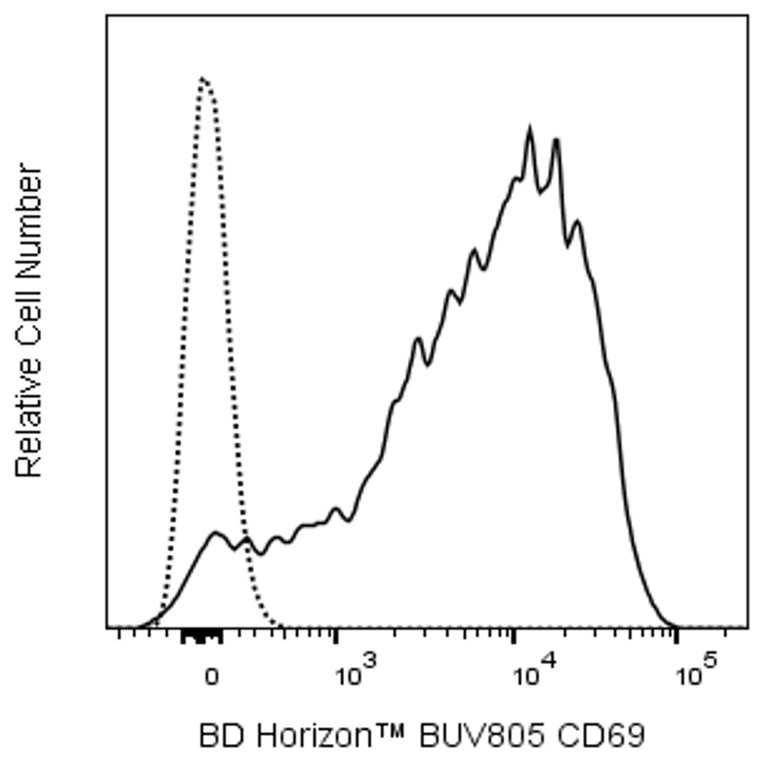
The FN50 monoclonal antibody specifically binds to human CD69. CD69 is also known as activation-induced molecule (AIM), early activation antigen (EA-1), very early activation antigen (VEA), C-type lectin domain family 2 member C (CLEC2C), MLR-3, GP32/28 and Leu-23. CD69 is a transmembrane type II homodimer receptor. CD69 is comprised of disulfide-linked, differentially glycosylated core protein subunits that are approximately 28 and 34 kDa in size. Each subunit contains a C-type lectin domain.  CD69 is expressed on activated T, B, and natural killer (NK) lymphocytes, thymocytes, neutrophils, eosinophils and platelets. In normal peripheral blood, a small and variable percentage of lymphocytes typically express detectable membrane CD69 antigen. Upon activation, CD69 antigen expression increases on lymphocytes. Peak CD69 expression generally occurs within 18 hours of activation, preceding the appearance of HLA-DR, IL-2Rα (CD25) and transferrin receptor (CD71). CD69 is highly expressed on the bright CD3+ subset of thymocytes. FN50 monoclonal antibody labels NK cells and most lymphocytes of the follicular mantle and perifollicular/interfollicular zone as well as germinal center T cells of lymph nodes and tonsils. Studies indicate that CD69 serves as a signaling receptor in the activation of a variety of cell types.

569163 Rev. 1
Format Details
Down Arrow Up Arrow
BUV805
The BD Horizon Brilliant™ Ultraviolet 805 (BUV805) Dye is part of the BD Horizon Brilliant™ Ultraviolet family of dyes. This tandem fluorochrome is comprised of a BUV395 donor with an excitation maximum (Ex Max) of 351-nm and an acceptor dye with an emission maximum (Em Max) at 803-nm. BUV805, driven by BD innovation, is designed to be excited by the ultraviolet laser (355 nm) and detected using an optical filter centered near 805-nm (e.g., a 820/60 or a 780/60 bandpass filter). Please ensure that your instrument’s configurations (lasers and optical filters) are appropriate for this dye.
altImg
BUV805
Ultraviolet 355 nm
351 nm
803 nm
569163 Rev.1
Citations & References
Down Arrow Up Arrow

Development References (10)

  1. Beiske K, AAS-Eng DA, Smeland EB, Sundan A, Marton PF, Funderud S. Immunohistochemical and functional characterization of the mAb A91 (FN 50)-reactive activation antigen (CD69) expressed on subsets of normal and neoplastic lymphoid cells. In: Knapp W. W. Knapp .. et al., ed. Leucocyte typing IV : white cell differentiation antigens. Oxford New York: Oxford University Press; 1989:436-439.
  2. CD69. In: Zola H. Leukocyte and stromal cell molecules : the CD markers. Hoboken, N.J.: Wiley-Liss; 2007:161.
  3. Knapp W. W. Knapp .. et al., ed. Leucocyte typing IV : white cell differentiation antigens. Oxford New York: Oxford University Press; 1989:1-1182.
  4. Lin G-X, Yang X, Hollemweguer E, et al. Cross-reactivity of CD antibodies in eight animal species. In: Mason D. David Mason .. et al., ed. Leucocyte typing VII : white cell differentiation antigens : proceedings of the Seventh International Workshop and Conference held in Harrogate, United Kingdom. Oxford: Oxford University Press; 2002:519-523.
  5. Schlossman SF. Stuart F. Schlossman .. et al., ed. Leucocyte typing V : white cell differentiation antigens : proceedings of the fifth international workshop and conference held in Boston, USA, 3-7 November, 1993. Oxford: Oxford University Press; 1995.
  6. Schwarting R, Biedobitek G, Stein H. Cluster report: CD69. In: Knapp W. W. Knapp .. et al., ed. Leucocyte typing IV : white cell differentiation antigens. Oxford New York: Oxford University Press; 1989:428-429.
  7. Stein H, Schwarting R, Niedobitek G, Dallenbach F. Activation Section Report. In: Knapp W. W. Knapp .. et al., ed. Leucocyte typing IV : white cell differentiation antigens. Oxford New York: Oxford University Press; 1989:387-398.
  8. Tomescu C, Chehimi J, Maino VC, Montaner LJ. NK cell lysis of HIV-1-infected autologous CD4 primary T cells: requirement for IFN-mediated NK activation by plasmacytoid dendritic cells. 2007; 179(4):2097-2104. (Clone-specific: Flow cytometry). View Reference
  9. Wang SR, Zhong N, Zhang XM, et al. OMIP 071: A 31-Parameter Flow Cytometry Panel for In-Depth Immunophenotyping of Human T-Cell Subsets Using Surface Markers.. Cytometry A. 2021; 99(3):273-277. (Clone-specific: Flow cytometry). View Reference
  10. Yoshino N, Ami Y, Terao K, Tashiro F, Honda M. Upgrading of flow cytometric analysis for absolute counts, cytokines and other antigenic molecules of cynomolgus monkeys (Macaca fascicularis) by using anti-human cross-reactive antibodies. Exp Anim. 2000; 49(2):97-110. (Clone-specific: Flow cytometry). View Reference
View All (10) View Less
569163 Rev. 1

Please refer to Support Documents for Quality Certificates


Global - Refer to manufacturer's instructions for use and related User Manuals and Technical data sheets before using this products as described


Comparisons, where applicable, are made against older BD Technology, manual methods or are general performance claims.  Comparisons are not made against non-BD technologies, unless otherwise noted.

For Research Use Only. Not for use in diagnostic or therapeutic procedures.