







Preparation And Storage


Product Notices
- This reagent has been pre-diluted for use at the recommended Volume per Test. We typically use 1 × 10^6 cells in a 100-µl experimental sample (a test).
- An isotype control should be used at the same concentration as the antibody of interest.
- Caution: Sodium azide yields highly toxic hydrazoic acid under acidic conditions. Dilute azide compounds in running water before discarding to avoid accumulation of potentially explosive deposits in plumbing.
- Source of all serum proteins is from USDA inspected abattoirs located in the United States.
- Alexa Fluor® 647 fluorochrome emission is collected at the same instrument settings as for allophycocyanin (APC).
- The Alexa Fluor®, Pacific Blue™, and Cascade Blue® dye antibody conjugates in this product are sold under license from Molecular Probes, Inc. for research use only, excluding use in combination with microarrays, or as analyte specific reagents. The Alexa Fluor® dyes (except for Alexa Fluor® 430), Pacific Blue™ dye, and Cascade Blue® dye are covered by pending and issued patents.
- Alexa Fluor® is a registered trademark of Molecular Probes, Inc., Eugene, OR.
- For fluorochrome spectra and suitable instrument settings, please refer to our Multicolor Flow Cytometry web page at www.bdbiosciences.com/colors.
- Alexa Fluor® is a registered trademark of Life Technologies Corporation.
- Please refer to http://regdocs.bd.com to access safety data sheets (SDS).
- Please refer to www.bdbiosciences.com/us/s/resources for technical protocols.
Companion Products
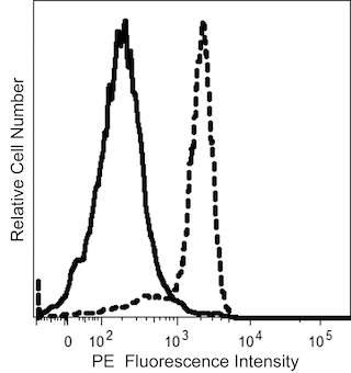


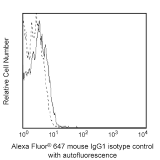
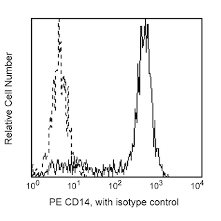
The 7-239 monoclonal antibody specifically binds to Sialic acid-binding Ig-like lectin 1 (Siglec-1), which is also known as Sialoadhesin (SN), or CD169. Siglec-1 is a type I transmembrane glycoprotein that belongs to the Siglec family within the Ig superfamily. This adhesion molecule especially binds to glycolipids and glycoproteins with terminal α-2 sialyl residues. Siglec-1 is expressed by macrophages and dendritic cells and serves as a cellular interaction molecule. Its expression can be upregulated by cells in response to type II collagen, or to cytokines including interferons, and tumor necrosis factor. Siglec-1 plays roles in endocytosis, hematopoiesis, and leucocyte migration. It mediates macrophage binding to various cell types including developing and mature leucocytes. Siglec-1 that is expressed by dendritic cells can also bind HIV-1 and may mediate viral transfer to bystander CD4+ T cells. Several Siglec-1 counter-receptors have been described including CD43, CD206, and CD227 which are expressed by T cells, macrophages, or breast cancer cells, respectively. The 7-239 antibody reportedly blocks Siglec-1 functions in some cellular assay systems.
Development References (6)
-
Hartnell A, Steel J, Turley H, Jones M, Jackson DG, Crocker PR. Characterization of human sialoadhesin, a sialic acid binding receptor expressed by resident and inflammatory macrophage populations. Blood. 2001; 97(1):288-296. (Biology). View Reference
-
Izquierdo-Useros N, Lorizate M, Puertas MC, et al. Siglec-1 is a novel dendritic cell receptor that mediates HIV-1 trans-infection through recognition of viral membrane gangliosides. PLoS ONE. 10(12)(Biology). View Reference
-
Kirchberger S, Majdic O, Steinberger P, et al. Human rhinoviruses inhibit the accessory function of dendritic cells by inducing sialoadhesin and B7-H1 expression. J Immunol. 2005; 175(2):1145-1152. (Immunogen: Blocking, Cell separation, Flow cytometry, Functional assay, Inhibition, Western blot). View Reference
-
Rose T, Grützkau A, Hirseland H, et al. IFNα and its response proteins, IP-10 and SIGLEC-1, are biomarkers of disease activity in systemic lupus rythematosus. Arthritis Rheum. 2013; 72(10):1639-1645. (Biology). View Reference
-
Xiong YS, Cheng Y, Lin QS, et al. Increased expression of Siglec-1 on peripheral blood monocytes and its role in mononuclear cell reactivity to autoantigen in rheumatoid arthritis. Rheumatology (Oxford). (Biology). View Reference
-
van den Berg TK, Nath D, Ziltener HJ, et al. Cutting edge: CD43 functions as a T cell counterreceptor for the macrophage adhesion eceptor sialoadhesin Siglec-1) . J Immunol. 2001; 166(6):3637-3640. (Biology). View Reference
Please refer to Support Documents for Quality Certificates
Global - Refer to manufacturer's instructions for use and related User Manuals and Technical data sheets before using this products as described
Comparisons, where applicable, are made against older BD Technology, manual methods or are general performance claims. Comparisons are not made against non-BD technologies, unless otherwise noted.
For Research Use Only. Not for use in diagnostic or therapeutic procedures.
