-
Your selected country is
Middle East / Africa
- Change country/language
Old Browser
This page has been recently translated and is available in French now.
Looks like you're visiting us from {countryName}.
Would you like to stay on the current country site or be switched to your country?
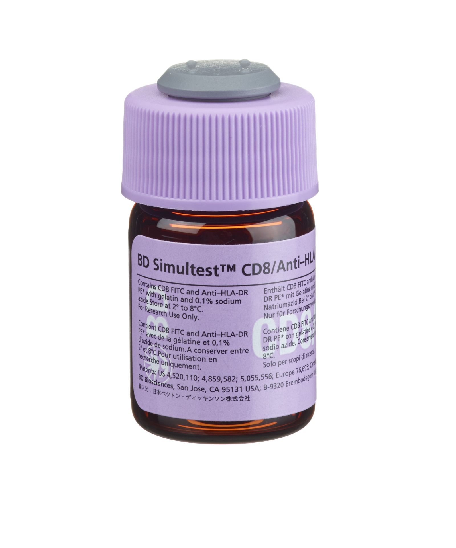
BD Simultest™ Anti-Human CD8 FITC/HLA-DR PE
(RUO (GMP))

Anti-Human CD8 FITC/HLA-DR PE
Regulatory Status Legend
Any use of products other than the permitted use without the express written authorization of Becton, Dickinson and Company is strictly prohibited.
Description
CD8, clone SK1, is derived from hybridization of mouse NS-1 myeloma cells with spleen cells from BALB/c mice immunized with human peripheral blood T lymphocytes. Anti-HLA-DR, clone L243, is derived from hybridization of mouse NS1/1-Ag4 myeloma cells with spleen cells from BALB/c mice immunized with the human lymphoblastoid B-cell line RPMI 8866. The CD8 (Leu-2a) antigen is present on the human suppressor/cytotoxic T-lymphocyte subset as well as on a subset of natural killer (NK) lymphocytes. The CD8 antigenic determinant interacts with class I major histocompatibility complex (MHC) molecules resulting in increased adhesion between the CD8+ T lymphocytes and the target cells. Binding of the CD8 antigen to class I MHC molecules enhances the activation of resting T lymphocytes. CD8 recognizes an antigen expressed on the 32-kdalton (kDa) α-subunit of a disulfide-linked bimolecular complex. The cytoplasmic domain of the α-subunit of the CD8 antigen is associated with the protein tyrosine kinase p56lck. The HLA-DR antigen is a human class II MHC antigen. The antigen is a transmembrane glycoprotein composed of α- and β-subunits that have molecular weights of 36 and 27 kDa, respectively. The antibody reacts with a nonpolymorphic HLA-DR epitope.
Preparation And Storage
Store vials at 2°C–8°C. Conjugated forms should not be frozen. Protect from exposure to light. Each reagent is stable until the expiration date shown on the bottle label when stored as directed.
| Description | Clone | Isotype | EntrezGene ID |
|---|---|---|---|
| CD8 FITC | SK1 | IgG1, κ | N/A |
| HLA-DR PE | L243 | IgG2a, κ | N/A |
Development References (36)
-
Anderson P, Blue ML, Morimoto C, Schlossman SF. Cross-linking of T3 (CD3) with T4 (CD4) enhances the proliferation of resting T lymphocytes. J Immunol. 1987; 139:678-682. (Biology).
-
Brodsky FM. A matrix approach to human class II histocompatibility antigens: reactions of four monoclonal antibodies with the products of nine haplotypes.. Immunogenetics. 1984; 19(3):179-94. (Biology). View Reference
-
Carney WP, Iacoviello V, Hirsh MS. Functional properties of T lymphocytes and their subsets in cytomegalovirus mononucleosis. J Immunol. 1983; 130:390-393. (Biology).
-
Centers for Disease Control. Update: universal precautions for prevention of transmission of human immunodeficiency virus, hepatitis B virus, and other bloodborne pathogens in healthcare settings. MMWR. 1988; 37:377-388. (Biology).
-
Clinical and Laboratory Standards Institute. 2005. (Biology).
-
Edwards JA, Durant BM, Jones DB, Evans PR, Smith JL. Differential expression of HLA class II antigens in fetal human spleen: relationship of HLA-DP, DQ, and DR to immunoglobulin expression.. J Immunol. 1986; 137(2):490-7. (Biology). View Reference
-
Eichmann K, Johnson J, Falk I, Emmrich F. Effective activation of resting mouse T lymphocytes by cross-linking submitogenic concentrations of the T-cell antigen receptor with either Lyt-2 or L3T4. Eur J Immunol. 1987; 17:643-650. (Biology).
-
Engleman EG, Warnke R, Fox RI, Dilley J, Benike CJ, Levy R. Studies of a human T lymphocyte antigen recognized by a monoclonal antibody.. Proc Natl Acad Sci USA. 1981; 78(3):1791-5. (Biology). View Reference
-
Evans RL, Wall DW, Platsoucas CD, et al. Thymus-dependent membrane antigens in man: inhibition of cell-mediated lympholysis by monoclonal antibodies to TH2 antigen. Proc Natl Acad Sci U S A. 1981; 78(1):544-548. (Biology). View Reference
-
Gallagher PF, Fazekas de St. Groth B, Miller JFAP. CD4 and CD8 molecules can physically associate with the same T-cell receptor. Proc Natl Acad Sci USA. 1989; 86:10044-10048. (Biology).
-
Giorgi J, Hultin L. Lymphocyte subset alterations and immunophenotyping by flow cytometry in HIV disease. Clin Immunol Newslett. 1990; 10(4):55-61. (Biology).
-
Giorgi JV, Detels R. T-cell subset alterations in HIV-infected homosexual men: NIAID multicenter AIDS cohort study. Clin Immunol Immunopathol. 1989; 52:10-18. (Biology).
-
Lampson LA, Levy R. Two populations of Ia-like molecules on a human B cell line.. J Immunol. 1980; 125(1):293-9. (Biology). View Reference
-
Landay A, Ohlsson-Wilhelm B, Giorgi JV. Application of flow cytometry to the study of HIV infection. AIDS. 1990; 4(6):479-497. (Biology). View Reference
-
Lanier LL, Le AM, Phillips JH, Warner NL, Babcock GF. Subpopulations of human natural killer cells defined by expression of the Leu-7 (HNK-1) and Leu-11 (NK-15) antigens. J Immunol. 1983; 131(4):1789-1796. (Biology). View Reference
-
Ledbetter JA, Evans RL, Lipinski M, Cunningham-Rundles C, Good RA, Herzenberg LA. Evolutionary conservation of surface molecules that distinguish T lymphocyte helper/inducer and cytotoxic/suppressor subpopulations in mouse and man. J Exp Med. 1981; 153(2):310-323. (Biology). View Reference
-
Linker-Israeli M, Gray J, Quismorio F, Horowitz D. Characterization of lymphocytes that suppress IL-2 production in systemic lypus erythematosus. Clin Exp Immunol. 1988; 73:236-241. (Biology).
-
Lum LG. The kinetics of immune reconstitution after human marrow transplantation. Blood. 1987; 69:369-380. (Biology).
-
Moebius U. Knapp W, Dörken B, Gilks W, et al, ed. Leucocyte Typing IV. White Cell Differentiation Antigens. New York: Oxford University Press; 1989:342-343.
-
Nicholson JKA, Jones BM. Immunophenotyping by flow cytometry: its use in HIV infection. Labmedica. 1989; 6:21-26. (Biology).
-
Pantaleo G, De Maria A, Koenig S, et al. CD8+ T lymphocytes of patients with AIDS maintain normal broad cytolytic function despite the loss of human immunodefieciency virus-specific cytotoxicity. Proc Natl Acad Sci USA. 1990; 87:4818-4822. (Biology).
-
Pantaleo G, Koenig S, Baseler M, Lane HC, Fauci AS. Defective clonogenic potential of CD8+ T lymphocytes in patients with AIDS. Expansion in vivo of a nonclonogenic CD3+CD8+DR+CD25- T cell population. J Immunol. 1990; 144:1696-1704. (Biology).
-
Pitzalis C, Kingsley G, Lanchbury J, Murphy J, Panayi G. Expression of HLA-DR, DQ and DP antigens and interleukin-2 receptor on synovial fluid T lymphocyte subsets in rheumatoid arthritis: Evidence for "frustrated" activation. J Rheumatol. 1987; 14:662-666. (Biology).
-
Prince HE, Arens L, Kleinman SH. CD4 and CD8 subsets defined by dual-color cytofluorometry which distinguish symptomatic from asymptomatic blood donors seropositive for human immunodeficiency virus. Diag Clin Immunol. 1987; 5:188-193. (Biology).
-
Reichert T, DeBruyere M, Deneys V, et al. Lymphocyte subset reference ranges in adult Caucasians. Clin Immunol Immunopathol. 1991; 60(2):190-208. (Biology). View Reference
-
Robbins PA, Evans EL, Ding AH, Warner NL, Brodsky FM. Monoclonal antibodies that distinguish between class II antigens (HLA-DP, DQ, and DR) in 14 haplotypes.. Hum Immunol. 1987; 18(4):301-13. (Biology). View Reference
-
Rudd CE, Burgess KE, Barber EK, Schlossman SF. Knapp W, Dörken B, Gilks WR, et al, ed. Leucocyte Typing IV: White Cell Differentiation Antigens. New York, NY: Oxford University Press; 1989:326-327.
-
Salazar-Gonzalez JF, Moody DJ, Giorgi JV, Martinez-Maza O, Mitsuyasu RT, Fahey JL. Reduced ecto-5'-nucleotidase activity and enhanced OKT10 and HLA-DR expression on CD8 (T suppressor/cytotoxic) lymphocytes in the acquired immune deficiency syndrome: evidence of CD8 cell immaturity. J Immunol. 1985; 135(3):1778-1785. (Biology). View Reference
-
Stites DP, Casavant CH, McHugh TM, et al. Flow cytometric analysis of lymphocyte phenotypes in AIDS using monoclonal antibodies and simultaneous dual immunofluorescence.. Clin Immunol Immunopathol. 1986; 38(2):161-77. (Biology). View Reference
-
Terstappen LW, Hollander Z, Meiners H, Loken MR. Quantitative comparison of myeloid antigens on five lineages of mature peripheral blood cells. J Leukoc Biol. 1990; 48(2):138-148. (Biology). View Reference
-
Tomkinson BE, Wagner DK, Nelson DL, Sullivan JL. Activated lymphocytes during acute Epstein-Barr virus infection.. J Immunol. 1987; 139(11):3802-7. (Biology). View Reference
-
Warnke R, Miller R, Grogan T, Pederson M, Dilley J, Levy R. Immunologic phenotype in 30 patients with diffuse large-cell lymphoma.. N Engl J Med. 1980; 303(6):293-300. (Biology). View Reference
-
Warnke RA, Levy R. Detection of T and B cell antigens with hybridoma monoclonal antibodies: a biotinavidin-horseradish peroxidase method. J Histochem Cytochem. 1980; 28:771-776. (Biology).
-
Wood GS, Warner NL, Warnke RA. Anti–Leu-3/T4 antibodies react with cells of monocyte/macrophage and Langerhans lineage. J Immunol. 1983; 131(1):212-216. (Biology). View Reference
-
Zipf TF, Fox RI, Dilley J, Levy R. Definition of the high-risk acute lymphoblastic leukemia patient by immunological phenotyping with monoclonal antibodies.. Cancer Res. 1981; 41(11 Pt 2):4786-9. (Biology). View Reference
-
van Es A, Baldwin WM, Oljans PJ, Tanke HJ, Ploem JS, van Es LA. Expression of HLA-DR on T lymphocytes following renal transplantation, and association with graft-rejection episodes and cytomegalovirus infection.. Transplantation. 1984; 37(1):65-9. (Biology). View Reference
Please refer to Support Documents for Quality Certificates
Global - Refer to manufacturer's instructions for use and related User Manuals and Technical data sheets before using this products as described
Comparisons, where applicable, are made against older BD Technology, manual methods or are general performance claims. Comparisons are not made against non-BD technologies, unless otherwise noted.
For Research Use Only. Not for use in diagnostic or therapeutic procedures.
Although not required, these products are manufactured in accordance with Good Manufacturing Practices.