Quick Guide to Memory T Cells
September 07, 2022
Introduction to memory T cells
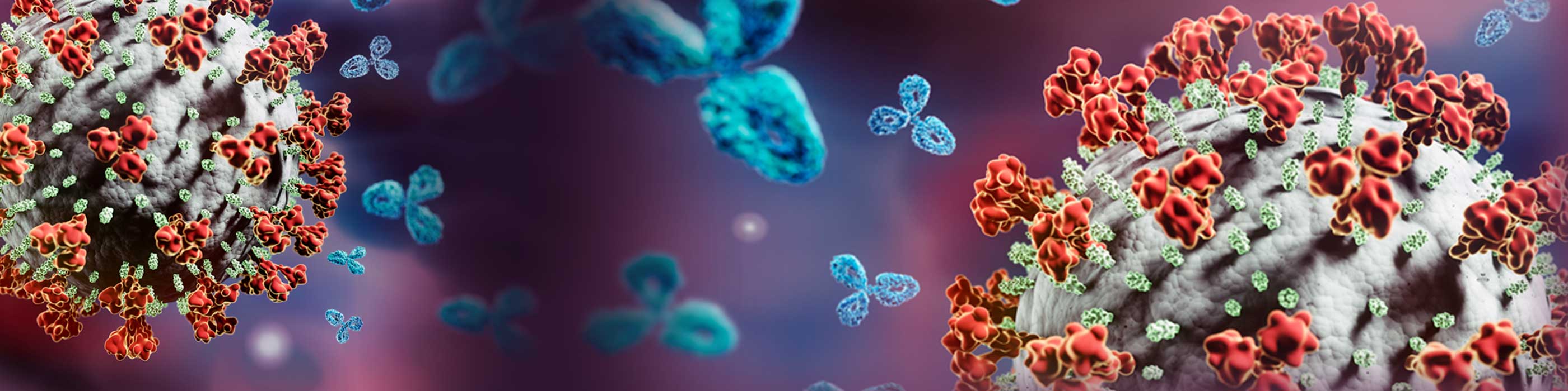
Antigen-specific memory T cells have provided highly efficient and long-lasting immunity against infections.1 The important role of T cell-mediated immunity in prophylactic vaccines has become more evident over the years, in diseases such as hepatitis C, human immunodeficiency virus (HIV) and cytomegalovirus (CMV).2
These antigen-specific T cell responses originate from a small number of naïve precursor cells, which, following several phases, are able to stably persist as memory T cells even in the absence of antigens.1 These patterns of diversification observed during infection or in response to vaccination are important for the quality of antigen-specific immunity.1
The stages of T cell diversification
- Antigen-specific interactions between T cells and dendritic cells can last for up to 12 hours, while CD4+ and CD8+ T cells receive necessary signals for activation.2
- This activation results in alteration of expression of various molecules, including integrins, selectins, and chemokine receptors, which, in turn promote T cell proliferation, differentiation, and migration of T cells to inflamed tissue.2
- Following resolution of the infection, 90-95% of the effector T cells are eliminated by programmed cell death, and a small but diverse pool of memory cells survive.2
- This led to the identification of two distinct subsets: effector memory T cells and central memory T cells, and more recently, tissue resident-memory T cells.2
Upon re-exposure to antigens in a secondary immune response, memory T cells undergo rapid population expansion and mediate more robust effector functions compared with the primary immune response, which leads to rapid clearance of the infection.3 Essential features of memory T cells are (1) evidence of prior expansion and/or activation, (2) persistence in the absence of antigens, (3) enhanced functional activity upon antigen re-exposure.3
The subsets: central, effector and tissue resident memory T cells
T cell subsets can then be further characterised based on their effector memory differentiation status. Every effector/memory subset has unique characteristics which provide them with differential migratory capacity, longevity, and functionality.
| Effector memory T cells | Central memory T cells | Tissue resident T cells (TRM) | |
|---|---|---|---|
| How can they be identified? | Combined expression and/or lack of cell surface markers (e.g., KLRG1hi/CD44hi/ CD127lo/CD62Llo)2 | Expression of KLRG1lo/ CD44hi/CD127hi/CD62hi surface markers2 | Expression of CD69hi/CD62Llo/CD44hi and other surface markers (e.g., CD11a, CD38, CD49a)2 |
| Where do they reside? | Non-lymphoid tissue and in peripheral circulation2 | Secondary lymphoid organs and in peripheral circulation2 | Most organs and peripheral tissue2 |
| Proliferation capacity? | Limited2 | Superior2 | Limited1 |
| Circulation capacity? | Yes2 | Yes2 | No2 |
Multiparametric T cell assays
Multiparametric assays, such as flow cytometry using fluorochrome-conjugated cell surface antigen-specific antibodies, can identify T cell activation markers and provide key insights into a range of research areas, including the development of new diagnostic and prognostic tests.1,4
For instance, the ability to observe the magnitude of a vaccine-elicited T cell response may serve as a predictor for efficacy in vaccination settings.2 Research has shown that the size of a CMV viral inoculation directly influences the degree of memory T cell inflation and degree of immune alterations in long-lasting infections; data show a clear correlation between the magnitude of the CD8+ T cell response and the frequency of these cells exhibiting an effector memory phenotype.5,6
Significant decreases have been observed in the counts of CD3+, CD4+, and CD8+ T cells in the peripheral blood of patients with COVID-19, more so in severe COVID-19 cases compared to mild-to-moderate illness.4 CD4+ and CD8+ were selected as candidate diagnostic markers in the diagnosis of COVID-19 and prediction of severe cases.4
In the treatment of some cancers, CD44 expression may act as a biomarker for worse prognosis. CD44 expression in the mucosa of patients with oral squamous cell carcinoma (OSCC) was significantly higher than in normal mucosa, and was higher in dysplastic than non-dysplastic leukoplakia. 7 This indicates a correlation between the expression this cell adhesion molecule as a cancer stem cell antigen and transition to OSCC.7
Useful resources for carrying out your flow cytometry experiment