
Apoptosis is an organized process that signals cells to self-destruct for cell renewal or to control aberrant cell growth. As cells become damaged or are no longer needed, they undergo apoptosis or programmed cell death, a normal physiological process that occurs during embryonic development and tissue homeostasis. Apoptosis controls the orderly death of damaged cells, whereas necrosis occurs as a result of tissue damage, causing the loss of both damaged and surrounding cells.
Apoptotic process
The apoptotic process is characterized by certain morphological features. These include changes in the plasma membrane (such as loss of membrane symmetry and attachment), a condensation of the cytoplasm and nucleus, protein cleavage, and internucleosomal cleavage of DNA. In the final stages of the process, dying cells become fragmented into apoptotic bodies and consequently are eliminated by phagocytic cells without significant inflammatory damage to surrounding cells.
However, some cell types do not display characteristic features of apoptosis. In those cases, multiple aspects of apoptosis might need to be analyzed to confirm the mechanism of cell death.
To support this spectrum of requirements, BD Biosciences offers a full range of apoptosis detection tools and technologies for measuring indicators at different stages across the apoptotic process. BD Biosciences tools use multiple methodologies including flow cytometry, bioimaging and microscopy (for live and fixed cell analysis) as well as ELISA, IHC, western blot and spectrofluorometry.


Methods for detecting apoptosis or dead cells (viability) by cell preparation type.
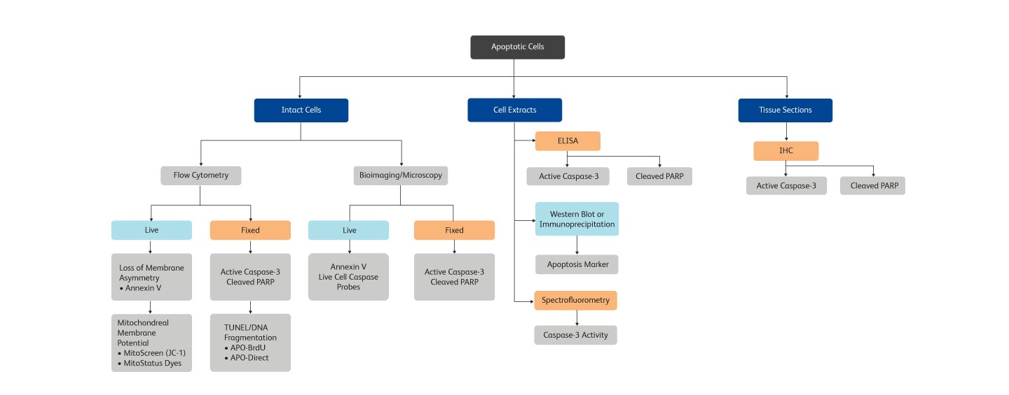
Methods for detecting apoptosis in different cell or tissue types
Annexin V—a key protein in apoptosis signaling
Changes in the plasma membrane are one of the characteristics of the apoptotic process detected in living cells. Apoptosis can be detected by the presence of phosphatidylserine (PS), which is normally located on the cytoplasmic face of the plasma membrane. During apoptosis, PS translocates to the outer leaflet of the plasma membrane and can be detected by flow cytometry and cell imaging through binding to fluorochrome-labeled annexin V when calcium is present.
BD Biosciences offers annexin V in several common formats such as FITC, PE, BD Horizon™ BV421 for the violet laser, and BD Horizon™ BUV395 for the ultraviolet laser. With the addition of these new formats, more complex assays can be developed to look at apoptosis within heterogeneous cell subsets.
Since intracellular annexin V is also exposed if the plasma membrane is compromised, a membrane-impermeant dye such as 7-AAD is commonly used to distinguish between apoptotic and dead cells to exclude the dead cells. The populations of cells that are stained with annexin V only represent the apoptotic cell populations.


Radio-frequency dose-dependent apoptosis, necrosis and cell death monitored by annexin V-BD Horizon™ V450. Radio-frequency dose-dependent apoptosis, necrosis and cell death monitored by annexin V-BD Horizon™ V450 in pancreatic carcinoma cell lines treated with a low dose of cetuximab targeted gold nanoparticles. As the RF field power increases, the temperature increases, and a shift from apoptosis (lower right quadrant) to frank necrosis (upper left quadrant) is seen.
Measurement of cleaved caspases and PARP
One of the most consistently observed characteristics of apoptosis is the activation of a series of cytosolic proteases called caspases. Caspases are activated upon cleavage at aspartate residues during the earliest stages of apoptosis. Active caspases can then cleave many proteins, including poly-ADP ribose polymerase (PARP), other caspases and other protein substrates en masse. This leads to the loss of cellular structure and function, and ultimately results in cell death. Caspases-9, -8 and -3 have been implicated in apoptosis—caspase-9 in the mitochondrial pathway, caspase-8 in the Fas/CD95 pathway, and caspase-3 more downstream, activated by multiple pathways.
BD Biosciences carries a variety of reagents to measure caspases, particularly caspase-3. They include antibodies directed exclusively against the active form of the caspase. These antibodies are available in a variety of formats and can be used for flow cytometry, imaging, ELISA and western blot.
BD Biosciences also offers a range of tools for caspase activity assays from individual fluorogenic peptide substrates and inhibitors, to kits, to ready-to-use assay plates. All are based on the use of synthetic tetrapeptide substrates that are designed such that proteolytic cleavage by active human or mouse caspases results in release of a fluorophore or chromophore. The individual synthetic tetrapeptide substrates, together with the caspase inhibitors and active caspase enzymes, offer flexibility in the experimental design of a caspase activity assay.

BD also offers reagents for the detection of caspase activity in intact, unfixed cells for flow cytometry in the form of live cell caspase probes. Because these probes are cell permeable, they do not require fixation of the target cells before use and can be incubated directly with the intact, unfixed samples to quickly detect caspase activity. These are available for three lasers for ease of panel design.
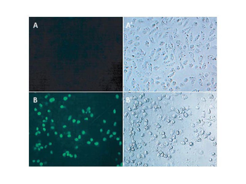
Flow cytometric analysis of apoptotic and nonapoptotic populations using anti-active caspase-3 antibodies. Jurkat T cells (A, A1) or mouse thymocytes (B, B1) were left untreated (A, B) or treated for 4 h with camptothecin (A1) or a mouse Fas monoclonal antibody, clone Jo2 (Cat. No. 554254) to induce apoptosis (B1). Cells were permeabilized and then stained with PE-conjugated active caspase-3 antibodies (Cat. No. 557091). Untreated cells were primarily negative for the presence of active caspase-3, whereas about half of each population of cells induced to undergo apoptosis had detectable active caspase-3.

Obtain the complete picture
In addition to caspases and annexin V, there are several other proteins important for the study of apoptosis, including the Bcl-2 family, tumor necrosis factor receptor (TNFR) family, PARP and other signaling molecules.
Bcl-2 family members, identified by the presence of conserved Bcl-2 homology (BH3) domains, are versatile key regulators of apoptosis. Bcl-2, for example, protects cells from apoptosis by associating with the mitochondrial membrane and preventing the release of cytochrome c from the mitochondria. In contrast other Bcl-2 family members, such as Bax, promote apoptosis. Increased levels of Bcl-2 have been reported in cancer.
The TNFR family contains many members, including CD95, that can be divided into three major groups based on structure. Signaling through the TNFR pathway leads to apoptosis.
PARPs are DNA repair enzymes that are activated by DNA strand breaks. Cleavage of PARP by caspase-3 into 24- and 89-kDa fragments inactivates the PARP enzyme.
BD Biosciences carries antibodies specific for cleavage products of PARP that are useful markers of apoptosis. These antibodies are available in a variety of formats and can be combined with other markers to gain additional information about the cell.
Other BD Biosciences tools to streamline apoptosis research
There are many apoptosis triggers including certain cytokines, protein-protein interactions and chemicals. Once apoptosis starts, changes in the mitochondria membrane potential can be measured by flow cytometry using the BD® MitoScreen (JC-1) Flow Cytometry Kit or the BD Pharmingen™ MitoStatus Dyes. Decreases in mitochondrial membrane potential lead to increased mitochondrial membrane permeability and the release of soluble proteins such as cytochrome c and pro-caspases.
DNA fragmentation is one of the last phases in apoptosis, resulting from the activation of endonucleases during the apoptotic process. There are several established methods for the study of DNA fragmentation including isolation and separation of DNA fragments by agarose gel electrophoresis and end labeling. The BD® APO-BrdU™ Kit uses end labeling or the terminal deoxynucleotidyl transferase (TdT) nick end labeling (TUNEL method) to support the study of DNA fragmentation. In this assay, TdT catalyzes a template-independent addition of bromolated deoxyuridine triphosphates (Br-dUTP) to the 3'-hydroxyl (OH) termini of double- and single-stranded DNA. After the Br-dUTP is incorporated, these terminal sites of double- and single-stranded DNA are identified using flow cytometry by staining cells with labeled anti-BrdU. In contrast, the BrdU proliferation assay incorporates BrdU into newly synthesized DNA, into sites of DNA strand breaks.
At the end of apoptosis, cells become non-viable. Differentiating apoptotic from necrotic or dead cells is therefore key to isolating apoptotic populations. BD offers a number of viability products for identifying dead cells, including esterase activity probes and membrane integrity dyes.
With an overwhelming number of available techniques and products, selecting the most appropriate method is often difficult. To help make this choice easier, the overview below summarizes commercially available assays from a biological perspective.
| Feature Measured | Assays | Key Features |
|---|---|---|
| Plasma membrane alterations (phosphatidylserine exposure) | Annexin binding assay Single conjugates Annexin V kits |
Detects early apoptosis markers Quick and easy Flow cytometry or immunofluorescence application |
| Caspase activation by spectrofluorometry | Caspase activity assay kits and reagents | Quick and easy |
| Caspase activation by immunoassay | Active caspase-3 immunoassays, including active caspase-3 conjugates | ELISA, flow cytometry or western blot in fixed cells or lysed tissues |
| Caspase activation by live cell activity probes | Active caspase-3 live cell caspase probes: BD Pharmingen™ Yellow-Green Live Cell Caspase Probe BD Pharmingen™ Blue Live Cell Caspase Probe BD Pharmingen™ Violet Live Cell Caspase Probe |
Flow cytometry Allows caspase activity detection in intact, unfixed cells |
| Key protein activity | Key signaling protein conjugates, including: Cleaved PARP Bcl-2 CD95 H2AX |
Available for flow cytometry, western blot, immunohistochemistry and immunofluorescence Available in multiple formats, which allows multiplexing with other apoptosis and viability tools for better characterization of populations of interest |
| Mitochondrial changes | BD® MitoScreen Kit BD Pharmingen™ MitoStatus TMRE BD Pharmingen™ MitoStatus Red |
Fast, easy, single-cell resolution by flow cytometry or fluorescent microscopy |
| DNA fragmentation | APO-BrdU™ TUNEL Assay APO-DIRECT™ TUNEL Assay |
Works with adherent cells, single-cell resolution in conjunction with cell cycle analysis by flow cytometry |
| Esterase activity | BD Pharmingen™ Calcein AM BD Pharmingen™ Calcein Blue AM |
Measures viability in intact, unfixed cells by flow cytometry |
| Membrane integrity by nucleic acid detection | Propidium Iodide 7-AAD DAPI DRAQ7™ BD Via-Probe™ Green Nucleic Acid Stain BD Via-Probe™ Red Nucleic Acid Stain |
Measures viability in intact, unfixed cells for flow cytometry by staining nucleic acid in dead cells, which are permeable to the dyes Available for multiple laser lines for ease of panel design |
| Membrane integrity by dye exclusion | BD Horizon™ Fixable Viability Stains | Measures viability in fixed cells for flow cytometry by staining the surface of intact cells and the surface and interior of dead cells Available in 10 colors for ease of panel design |
Simultaneous studies of apoptosis, cell cycle and DNA damage
Apoptosis and cell proliferation assays are particularly useful for basic cancer research and drug discovery. Comparing data across different experiments can be challenging due to variability introduced by sample handling, timing and variability within the sample.
Multicolor flow cytometry addresses these challenges and is an excellent tool to study apoptosis and cell proliferation. Relevant markers can be combined with cell phenotyping markers to look at events within subpopulations of cells. Antibodies to phosphoproteins can be used to examine phosphorylation events.

-
Application Notes

-
Brochure
-
Product Information Sheets
-
Protocols
-
 Annexin V with Cell Surface Antibody Staining for Suspension Cells
-
Annexin V Staining of Adherent Cells for Flow Cytometry
-
Annexin V Staining Protocol
-
APO-BrdU™ Procedure (TUNEL Assay for Flow)
-
APO-DIRECT™ Procedure (TUNEL Assay for Flow)
-
Cell Death Induced by Camptothecin
-
Cell Death Induced by Staurosporine
-
Induction of Apoptosis Using Anti-Human Fas (CD95), Clone DX2
-
Induction of Apoptosis Using Anti-Mouse Fas (CD95), Clone Jo2
-
Webinars
For Research Use Only. Not for use in diagnostic or therapeutic procedures.
Alexa Fluor is a trademark of Life Technologies Corporation. APO-BRDU and APO-DIRECT are trademarks of Phoenix Flow Systems. DRAQ7 is a trademark of BioStatus Ltd. Triton is a trademark of Dow Chemical Company.