







Preparation And Storage


Product Notices
- Since applications vary, each investigator should titrate the reagent to obtain optimal results.
- An isotype control should be used at the same concentration as the antibody of interest.
- Caution: Sodium azide yields highly toxic hydrazoic acid under acidic conditions. Dilute azide compounds in running water before discarding to avoid accumulation of potentially explosive deposits in plumbing.
- Sodium azide is a reversible inhibitor of oxidative metabolism; therefore, antibody preparations containing this preservative agent must not be used in cell cultures nor injected into animals. Sodium azide may be removed by washing stained cells or plate-bound antibody or dialyzing soluble antibody in sodium azide-free buffer. Since endotoxin may also affect the results of functional studies, we recommend the NA/LE (No Azide/Low Endotoxin) antibody format, if available, for in vitro and in vivo use.
- Please refer to www.bdbiosciences.com/us/s/resources for technical protocols.
Companion Products



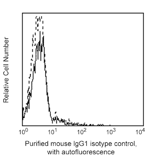
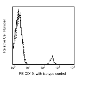
The 3A2-2E7 monoclonal antibody (also known as SN8) specifically recognizes CD79b. Immunoglobulin (Ig) antigen receptors are composed of a non-covalently-associated complex of Ig and two other proteins, Igα and Igβ, clustered as CD79a and CD79b, respectively. CD79b is a membrane glycoprotein of 229 residues, with a predicted relative molecular mass of 36-40 kDa. Its expression is restricted to B lineage cells. CD79b reportedly associates with surface IgM and is involved in signal transduction. The 3A2-2E7 antibody has similar reactivity characteristics as clone CB3-1. The 3A2-2E7 and CD3-1 antibodies specifically react with an epitope that is enhanced on certain B-cell leukemias such as prolymphocytic leukemia and lymphoma, but not on chronic lymphocytic leukemia.
Development References (7)
-
Astsaturov IA, Matutes E, Morilla R, et al. Differential expression of B29 (CD79b) and mb-1 (CD79a) proteins in acute lymphoblastic leukaemia. Leukemia. 1996; 10(5):769-773. (Biology). View Reference
-
Cragg MS, Chan HT, Fox MD, et al. The alternative transcript of CD79b is overexpressed in B-CLL and inhibits signaling for apoptosis. Blood. 2002; 100(9):3068-3076. (Biology). View Reference
-
Moreau EJ, Matutes E, A'Hern RP, et al. Improvement of the chronic lymphocytic leukemia scoring system with the monoclonal antibody SN8 (CD79b). Am J Clin Pathol. 1997; 108(4):378-382. (Biology). View Reference
-
Okazaki M, Luo Y, Han T, Yoshida M, Seon BK. Three new monoclonal antibodies that define a unique antigen associated with prolymphocytic leukemia/non-Hodgkin's lymphoma and are effectively internalized after binding to the cell surface antigen. Blood. 1993; 81(1):84-94. (Biology). View Reference
-
Schlossman SF. Stuart F. Schlossman .. et al., ed. Leucocyte typing V : white cell differentiation antigens : proceedings of the fifth international workshop and conference held in Boston, USA, 3-7 November, 1993. Oxford: Oxford University Press; 1995.
-
Van Kooten C, Galibert L, Seon BK, Garrone P, Liu YJ, Banchereau J. Cross-linking of antigen receptor via Ig-beta (B29, CD79b) can induce both positive and negative signals in CD40-activated human B cells. Clin Exp Immunol. 1997; 110(3):509-515. (Biology). View Reference
-
Zomas AP, Matutes E, Morilla R, Owusu-Ankomah K, Seon BK, Catovsky D. Expression of the immunoglobulin-associated protein B29 in B cell disorders with the monoclonal antibody SN8 (CD79b). Leukemia. 1996; 10(12):1966-1970. (Biology). View Reference
Please refer to Support Documents for Quality Certificates
Global - Refer to manufacturer's instructions for use and related User Manuals and Technical data sheets before using this products as described
Comparisons, where applicable, are made against older BD Technology, manual methods or are general performance claims. Comparisons are not made against non-BD technologies, unless otherwise noted.
For Research Use Only. Not for use in diagnostic or therapeutic procedures.
