Old Browser
Looks like you're visiting us from {countryName}.
Would you like to stay on the current country site or be switched to your country?


.png)

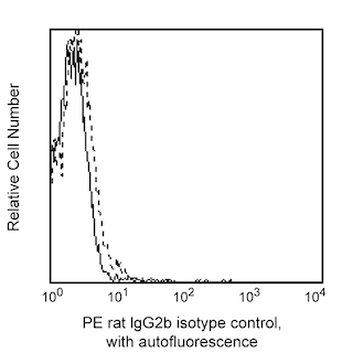
Flow cytometric analysis of TIM-2 expression on activated mouse B cells. BALB/c mouse splenocytes were stimulated in culture for 3 days with Purified NA/LE Hamster Anti-Mouse CD40 antibody (Cat. No. 553721) alone or in combination with either Recombinant Mouse IL-4 protein (Cat. No. 550067) or Donkey Anti-Mouse IgM F(ab')2 (Jackson ImmunoResearch Labs Cat. No. 715-006-020) as indicated. The cells were harvested, preincubated with BD Pharmingen™ Purified Rat Anti-Mouse CD16/CD32 (Mouse BD Fc Block™) (Cat. No. 553141/553142), and stained with Alexa Fluor® 647 Rat Anti-CD45R/B220 antibody (Cat. No. 557683) and either PE Rat IgG2b, κ Isotype Control (Cat. No. 555848; dotted line histograms) or PE Rat Anti-Mouse TIM-2 antibody (Cat. No. 566591; solid line histograms) at 0.5 μg/test. BD Via-Probe™ Cell Viability 7-AAD Solution (Cat. No. 555815/555816) was added to cells right before analysis. The histograms showing TIM-2 expression (or Ig Isotype Control staining) were derived from CD45R/B220-positive and 7-AAD-negative gated events with the with the forward and side light-scatter characteristics of viable leucocytes. Flow cytometric analysis was performed using a BD LSRFortessa™ Cell Analyzer System. Data shown on this Technical Data Sheet are not lot specific.
.png)

BD Pharmingen™ PE Rat Anti-Mouse TIM-2
.png)
Regulatory Status Legend
Any use of products other than the permitted use without the express written authorization of Becton, Dickinson and Company is strictly prohibited.
Preparation And Storage
Product Notices
- Since applications vary, each investigator should titrate the reagent to obtain optimal results.
- An isotype control should be used at the same concentration as the antibody of interest.
- Caution: Sodium azide yields highly toxic hydrazoic acid under acidic conditions. Dilute azide compounds in running water before discarding to avoid accumulation of potentially explosive deposits in plumbing.
- For fluorochrome spectra and suitable instrument settings, please refer to our Multicolor Flow Cytometry web page at www.bdbiosciences.com/colors.
- Please refer to www.bdbiosciences.com/us/s/resources for technical protocols.
Companion Products


The RMT2-26 monoclonal antibody specifically recognizes T cell immunoglobulin and mucin domain-2 (TIM-2) which is also known as T-cell membrane protein 2, TIMD-2, or Tim2. TIM-2 is an ~55 kDa type I transmembrane glycoprotein that is encoded by Timd2 (T cell immunoglobulin and mucin domain containing 2) which belongs to the immunoglobulin gene superfamily. TIM-2 contains extracellular IgV and mucin domains and a cytoplasmic region with a conserved tyrosine phosphorylation motif which is involved in transmembrane signaling. A human ortholog for mouse TIM-2 has not been identified. TIM-2 is reportedly expressed on B cells with higher levels on activated B cells and germinal center B cells, type-2 T helper (Th2) cells, oligodendrocytes, and epithelial cells in the kidney and liver. It may serve as a receptor for Semaphorin 4A (Sema4A) or the heavy chain of ferritin (H-ferritin). The RMT2-26 antibody can reportedly block the binding of H-ferritin to TIM-2.

Development References (8)
-
Abe Y, Kamachi F, Kawamoto T, et al. TIM-4 has dual function in the induction and effector phases of murine arthritis.. J Immunol. 2013; 191(9):4562-72. (Clone-specific: Flow cytometry). View Reference
-
Chakravarti S, Sabatos CA, Xiao S, et al. Tim-2 regulates T helper type 2 responses and autoimmunity.. J Exp Med. 2005; 202(3):437-44. (Biology). View Reference
-
Chen TT, Li L, Chung DH, et al. TIM-2 is expressed on B cells and in liver and kidney and is a receptor for H-ferritin endocytosis.. J Exp Med. 2005; 202(7):955-65. (Biology). View Reference
-
Han J, Seaman WE, Di X, et al. Iron uptake mediated by binding of H-ferritin to the TIM-2 receptor in mouse cells.. PLoS ONE. 2011; 6(8):e23800. (Biology). View Reference
-
Kawamoto T, Abe Y, Ito J, et al. Anti-T cell immunoglobulin and mucin domain-2 monoclonal antibody exacerbates collagen-induced arthritis by stimulating B cells.. Arthritis Res Ther. 2011; 13(2):R47. (Immunogen: Blocking, Flow cytometry, Immunoprecipitation, Western blot). View Reference
-
Kumanogoh A, Marukawa S, Suzuki K, et al.. Class IV semaphorin Sema4A enhances T-cell activation and interacts with Tim-2. Nature. 2002; 419(6907):629-633. (Biology). View Reference
-
Rennert PD, Ichimura T, Sizing ID, et al. T cell, Ig domain, mucin domain-2 gene-deficient mice reveal a novel mechanism for the regulation of Th2 immune responses and airway inflammation.. J Immunol. 2006; 177(7):4311-21. (Biology). View Reference
-
Rodriguez-Manzanet R, DeKruyff R, Kuchroo VK, Umetsu DT. The costimulatory role of TIM molecules. Immunol Rev. 2009; 229(1):259-270. (Biology). View Reference
Please refer to Support Documents for Quality Certificates
Global - Refer to manufacturer's instructions for use and related User Manuals and Technical data sheets before using this products as described
Comparisons, where applicable, are made against older BD Technology, manual methods or are general performance claims. Comparisons are not made against non-BD technologies, unless otherwise noted.
For Research Use Only. Not for use in diagnostic or therapeutic procedures.
Report a Site Issue
This form is intended to help us improve our website experience. For other support, please visit our Contact Us page.