The site is undergoing maintenance on
Thursday, September 10, 2026, 1:00 - 9:00 pm (BRT).
If you encounter problems while ordering, please contact us at BDBCustomerService@bd.com
See More
The site is undergoing maintenance on
Thursday, September 10, 2026, 1:00 - 9:00 pm (BRT).
If you encounter problems while ordering, please contact us at BDBCustomerService@bd.com
See MoreThe site is undergoing maintenance on
Thursday, September 10, 2026, 1:00 - 9:00 pm (BRT)
.If you encounter problems while ordering, please contact us at BDBCustomerService@bd.com
Looks like you're visiting us from {countryName}.
Would you like to stay on the current country site or be switched to your country?
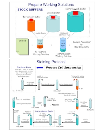
Two-color flow cytometric analysis of ZBTB7B (Th-POK) expression in human peripheral blood mononuclear cells. PBMCs were surface stained with Alexa Fluor® 488 Mouse Anti-Human CD4 antibody (Cat. No. 557695). The cells were then washed, fixed, and permeabilized using the BD Pharmingen™ Transcription Factor Buffer Set (Cat. No. 562574/562725) and stained with either PE Mouse IgG1, κ Isotype Control (Cat. No. 554680) or PE Mouse Anti-Human ZBTB7B (ThPOK) antibody. The two-color flow cytometric contour plots showing the correlated expression of ZBTB7B (ThPOK) [or Ig Isotype Control] versus CD4 were derived from gated events with the forward and side light-scatter characteristics of intact lymphocytes. Flow cytometric analysis was performed using a BD LSR Fortessa™ X-20 Flow Cytometer System.
Any use of products other than the permitted use without the express written authorization of Becton, Dickinson and Company is strictly prohibited.
The 6/hcKrox monoclonal antibody specifically recognizes human Th Inducing POZ-Kruppel Factor (ThPOK) which is also known as, cKrox or hcKrox. ThPOK is encoded by ZBTB7B (Zinc finger and BTB domain-containing protein 7B) and is a member of the POK/ZBT family of transcription regulators that play roles in lymphocyte development and the regulation of type I collagen genes. ThPOK expression is upregulated during the differentiation of CD4+ T helper cells but not CD8+ T cells. It indirectly increases the expression of CD4 in developing T cells by antagonizing Runx-3-mediated CD4 repression. ThPOK is also expressed in NKT and γδ T cells.

Please refer to Support Documents for Quality Certificates
Global - Refer to manufacturer's instructions for use and related User Manuals and Technical data sheets before using this products as described
Comparisons, where applicable, are made against older BD Technology, manual methods or are general performance claims. Comparisons are not made against non-BD technologies, unless otherwise noted.
For Research Use Only. Not for use in diagnostic or therapeutic procedures.