





BD Pharmingen™ PE Mouse Anti-Human Transferrin R (CD71)
Clone OKT9 (also known as OKT-9; OKT 9)
(RUO)


.png)


Preparation And Storage


Product Notices
- This reagent has been pre-diluted for use at the recommended Volume per Test. We typically use 1 × 10^6 cells in a 100-µl experimental sample (a test).
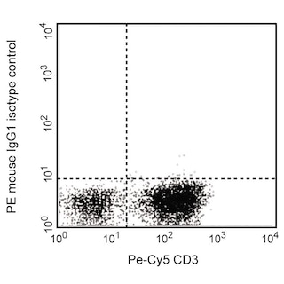
- An isotype control should be used at the same concentration as the antibody of interest.
- Source of all serum proteins is from USDA inspected abattoirs located in the United States.
- Caution: Sodium azide yields highly toxic hydrazoic acid under acidic conditions. Dilute azide compounds in running water before discarding to avoid accumulation of potentially explosive deposits in plumbing.
- For fluorochrome spectra and suitable instrument settings, please refer to our Multicolor Flow Cytometry web page at www.bdbiosciences.com/colors.
- Please refer to www.bdbiosciences.com/us/s/resources for technical protocols.
Companion Products
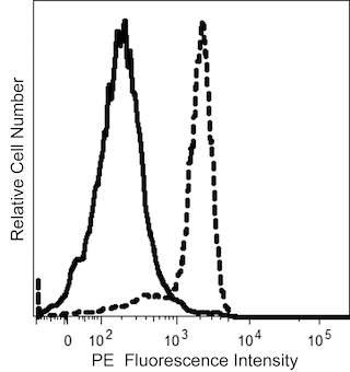


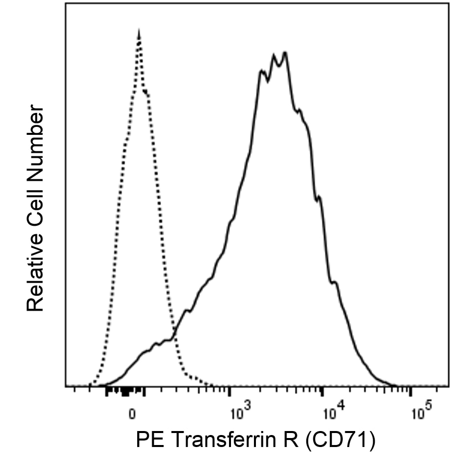
The OKT9 monoclonal antibody specifically binds to CD71 which is also known as the transferrin receptor (TFR or Transferrin R). CD71 (Transferrin R) is a type II transmembrane glycoprotein that is encoded by TFRC (transferrin receptor) which belongs to the Transferrin receptor family. It is expressed on cells as a disulfide-linked homodimer comprised of ~95 kDa monomers. CD71 is expressed on early thymocytes as well as activated lymphocytes, monocytes, macrophages, brain endothelium, and most proliferating cells. The transferrin receptor is also present on early erythroid cells but is lost as reticulocytes differentiate into mature erythrocytes. CD71 is lowly expressed on normal resting lymphocytes and is upregulated during lymphocyte responses to antigens or mitogens. The transferrin receptor is essential for iron transport into proliferating cells. Through an endocytic pathway, the transferrin receptor mediates cellular iron uptake by binding and internalizing iron that is bound to transferrin. After releasing iron within the low pH endosomal environment, transferrin and its receptor can be recycled to the cell surface.

Development References (4)
-
Goding JW, Burns GF. Monoclonal antibody OKT-9 recognizes the receptor for transferrin on human acute lymphocytic leukemia cells. J Immunol. 1981; 127(3):1256-1258. (Clone-specific: Immunoprecipitation). View Reference
-
Reinherz EL, Kung PC, Goldstein G, Levey RH, Schlossman SF. Discrete stages of human intrathymic differentiation: analysis of normal thymocytes and leukemic lymphoblasts of T-cell lineage. Proc Natl Acad Sci U S A. 1980; 77(3):1588-1592. (Immunogen: Flow cytometry). View Reference
-
Schwarting R, Stein H. Cluster report: CD71. In: Knapp W. W. Knapp .. et al., ed. Leucocyte typing IV : white cell differentiation antigens. Oxford New York: Oxford University Press; 1989:455-460.
-
Zola H, Swart B, Nicholson I, Voss E. CD71. In: Zola H. Leukocyte and Stromal Cell Molecules : the CD Markers. Hoboken, N.J.: Wiley-Liss; 2007:163.
Please refer to Support Documents for Quality Certificates
Global - Refer to manufacturer's instructions for use and related User Manuals and Technical data sheets before using this products as described
Comparisons, where applicable, are made against older BD Technology, manual methods or are general performance claims. Comparisons are not made against non-BD technologies, unless otherwise noted.
For Research Use Only. Not for use in diagnostic or therapeutic procedures.
