Preparation And Storage
Product Notices
- This reagent has been pre-diluted for use at the recommended Volume per Test. We typically use 1 × 10^6 cells in a 100-µl experimental sample (a test).
- An isotype control should be used at the same concentration as the antibody of interest.
- Caution: Sodium azide yields highly toxic hydrazoic acid under acidic conditions. Dilute azide compounds in running water before discarding to avoid accumulation of potentially explosive deposits in plumbing.
- Source of all serum proteins is from USDA inspected abattoirs located in the United States.
- Please observe the following precautions: Absorption of visible light can significantly alter the energy transfer occurring in any tandem fluorochrome conjugate; therefore, we recommend that special precautions be taken (such as wrapping vials, tubes, or racks in aluminum foil) to prevent exposure of conjugated reagents, including cells stained with those reagents, to room illumination.
- When excited by the yellow-green (561-nm) laser, the fluorescence may be brighter than when excited by the blue (488-nm) laser.
- Because of the broad absorption spectrum of the tandem fluorochrome, extra care must be taken when using multi-laser cytometers, which may directly excite both PE and CF™594.
- Texas Red is a registered trademark of Molecular Probes, Inc., Eugene, OR.
- CF™ is a trademark of Biotium, Inc.
- This product is provided under an Agreement between BIOTIUM and BD Biosciences. The manufacture, use, sale, offer for sale, or import of this product is subject to one or more patents or pending applications owned or licensed by Biotium, Inc. This product, and only in the amount purchased by buyer, may be used solely for buyer’s own internal research, in a manner consistent with the accompanying product literature. No other right to use, sell or otherwise transfer (a) this product, or (b) its components is hereby granted expressly, by implication or by estoppel. This product is for research use only. Diagnostic uses require a separate license from Biotium, Inc. For information on purchasing a license to this product including for purposes other than research, contact Biotium, Inc., 3159 Corporate Place, Hayward, CA 94545, Tel: (510) 265-1027. Fax: (510) 265-1352. Email: btinfo@biotium.com.
- For fluorochrome spectra and suitable instrument settings, please refer to our Multicolor Flow Cytometry web page at www.bdbiosciences.com/colors.
- Please refer to www.bdbiosciences.com/us/s/resources for technical protocols.
Companion Products


.png?imwidth=320)
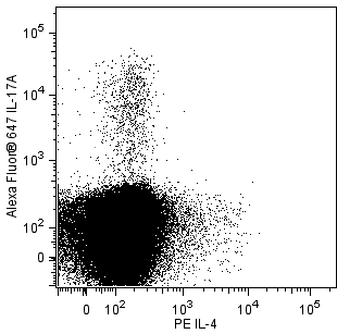
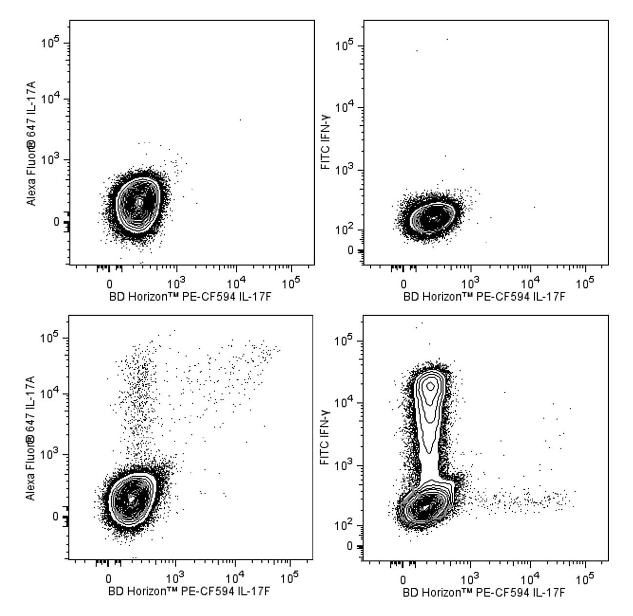
The O33-782 monoclonal antibody specifically binds to Interleukin-17F (IL-17F). IL-17F is a member of the IL-17 family of cytokines. IL-17F is encoded by the IL17F gene located in chromosome 6 (location: 6p12). IL-17F is a proinflammatory cytokine that is produced by activated T cells including differentiated CD4+ T helper 17 (Th17) cells. Activated Th17 cells can express disulfide-linked IL-17F and IL-17A homodimers as well as IL-17A/IL-17F heterodimers. These IL-17 dimers act by binding to and signaling through IL-17 receptor complexes (IL-17R). IL-17R are comprised of transmembrane IL-17RA and IL-17-RC protein subunits that are expressed by a variety of target cells including epithelial and endothelial cells, keratinocytes, fibroblasts, and granulocytes. IL-17F can induce target cells to produce proinflammatory cytokines such as IL-1β, IL-6, G-CSF, GM-CSF, and TNF and chemokines including CXCL1/Gro-α, CXCL2/Gro-β, and CXCL8/IL-8 that attract and activate leukocytes, eg, neutrophils. Th17 and other IL-17F-producing cells play protective roles in the clearance of extracellular pathogens, including bacteria and fungi. IL-17F can also play adverse roles in inflammation associated with asthma and autoimmune diseases.
This antibody is conjugated to BD Horizon™ PE-CF594, which has been developed exclusively by BD Biosciences as a better alternative to PE-Texas Red®. PE-CF594 excites and emits at similar wavelengths to PE-Texas Red® yet exhibits improved brightness and spectral characteristics. Due to PE having maximal absorption peaks at 496 nm and 564 nm, PE-CF594 can be excited by the blue (488-nm), green (532-nm) and yellow-green (561-nm) lasers and can be detected with the same filter set as PE-Texas Red® (eg 610/20-nm filter).

Development References (7)
-
Fouser LA, Wright JF, Dunussi-Joannopoulos K, Collins M. Th17 cytokines and their emerging roles in inflammation and autoimmunity. Immunol Rev. 2008; 226:87-102. (Biology). View Reference
-
Melton AC, Melrose J, Alajoki L, et al. Regulation of IL-17A production is distinct from IL-17F in a primary human cell co-culture model of T cell-mediated B cell activation. PLoS ONE. 2013; 8(3):e58966. (Clone-specific: Flow cytometry). View Reference
-
Shen F, Gaffen SL. Structure-function relationships in the IL-17 receptor: implications for signal transduction and therapy. Cytokine. 2008; 41(2):92-104. (Biology). View Reference
-
Starnes T, Robertson MJ, Sledge G, et al.. Cutting edge: IL-17F, a novel cytokine selectively expressed in activated T cells and monocytes, regulates angiogenesis and endothelial cell cytokine production. J Immunol. 2001; 167(8):4137-4140. (Biology). View Reference
-
Wang YH, Liu YJ. The IL-17 cytokine family and their role in allergic inflammation. Curr Opin Immunol. 2008; 20(6):697-702. (Biology). View Reference
-
Wright JF, Bennett F, Li B, et al. The human IL-17F/IL-17A heterodimeric cytokine signals through the IL-17RA/IL-17RC receptor complex. J Immunol. 2008; 181(4):2799-2805. (Biology). View Reference
-
Wright JF, Guo Y, Quazi A, et al. Identification of an interleukin 17F/17A heterodimer in activated human CD4+ T cells. J Biol Chem. 2007; 282(18):13447-13455. (Biology). View Reference
Please refer to Support Documents for Quality Certificates
Global - Refer to manufacturer's instructions for use and related User Manuals and Technical data sheets before using this products as described
Comparisons, where applicable, are made against older BD Technology, manual methods or are general performance claims. Comparisons are not made against non-BD technologies, unless otherwise noted.
For Research Use Only. Not for use in diagnostic or therapeutic procedures.