







Preparation And Storage


Recommended Assay Procedures
For optimal and reproducible results, BD Horizon Brilliant Stain Buffer should be used anytime two or more BD Horizon Brilliant dyes (including BD Optibuild Brilliant reagents) are used in the same experiment. Fluorescent dye interactions may cause staining artifacts which may affect data interpretation. The BD Horizon Brilliant Stain Buffer was designed to minimize these interactions. More information can be found in the Technical Data Sheet of the BD Horizon Brilliant Stain Buffer (Cat. No. 563794/566349) or the BD Horizon Brilliant Stain Buffer Plus (Cat. No. 566385).
Product Notices
- This reagent has been pre-diluted for use at the recommended Volume per Test. We typically use 1 × 10^6 cells in a 100-µl experimental sample (a test).
- An isotype control should be used at the same concentration as the antibody of interest.
- Caution: Sodium azide yields highly toxic hydrazoic acid under acidic conditions. Dilute azide compounds in running water before discarding to avoid accumulation of potentially explosive deposits in plumbing.
- Source of all serum proteins is from USDA inspected abattoirs located in the United States.
- Alexa Fluor® is a registered trademark of Molecular Probes, Inc., Eugene, OR.
- For fluorochrome spectra and suitable instrument settings, please refer to our Multicolor Flow Cytometry web page at www.bdbiosciences.com/colors.
- Cy is a trademark of GE Healthcare.
- BD Horizon Brilliant Stain Buffer is covered by one or more of the following US patents: 8,110,673; 8,158,444; 8,575,303; 8,354,239.
- BD Horizon Brilliant Violet 711 is covered by one or more of the following US patents: 8,110,673; 8,158,444; 8,227,187; 8,455,613; 8,575,303; 8,354,239.
- Please refer to http://regdocs.bd.com to access safety data sheets (SDS).
- Please refer to www.bdbiosciences.com/us/s/resources for technical protocols.
Companion Products




The DX11 monoclonal antibody specifically binds to CD226 which is also known as DNAX accessory molecule-1 (DNAM-1), Platelet and T cell activation antigen 1 (PTA1), or T lineage-specific activation antigen 1 antigen (TLiSA1). CD226 is a 65 kDa type 1 transmembrane glycoprotein consisting of 318 amino acid residues including two Ig-like domains. CD226 is expressed on the majority of T cells, NK cells, monocytes, platelets, and a subset of B cells, but not on erythrocytes. It is also present on a subset of thymocytes coexpressing high density surface CD3. CD226 is not present on normal fibroblast cell lines or tumor cell lines of epithelial or neuronal origins. CD226 is a tyrosine phosphorylated, signal-transducing molecule which participates in primary adhesion during cytotoxic T lymphocyte (CTL)- or NK cell-mediated cytotoxicity. The DX11 antibody inhibits T- and NK cell-mediated cytotoxicity against a variety of tumor cell targets, and blocks cytokine production by alloantigen-specific T cells.
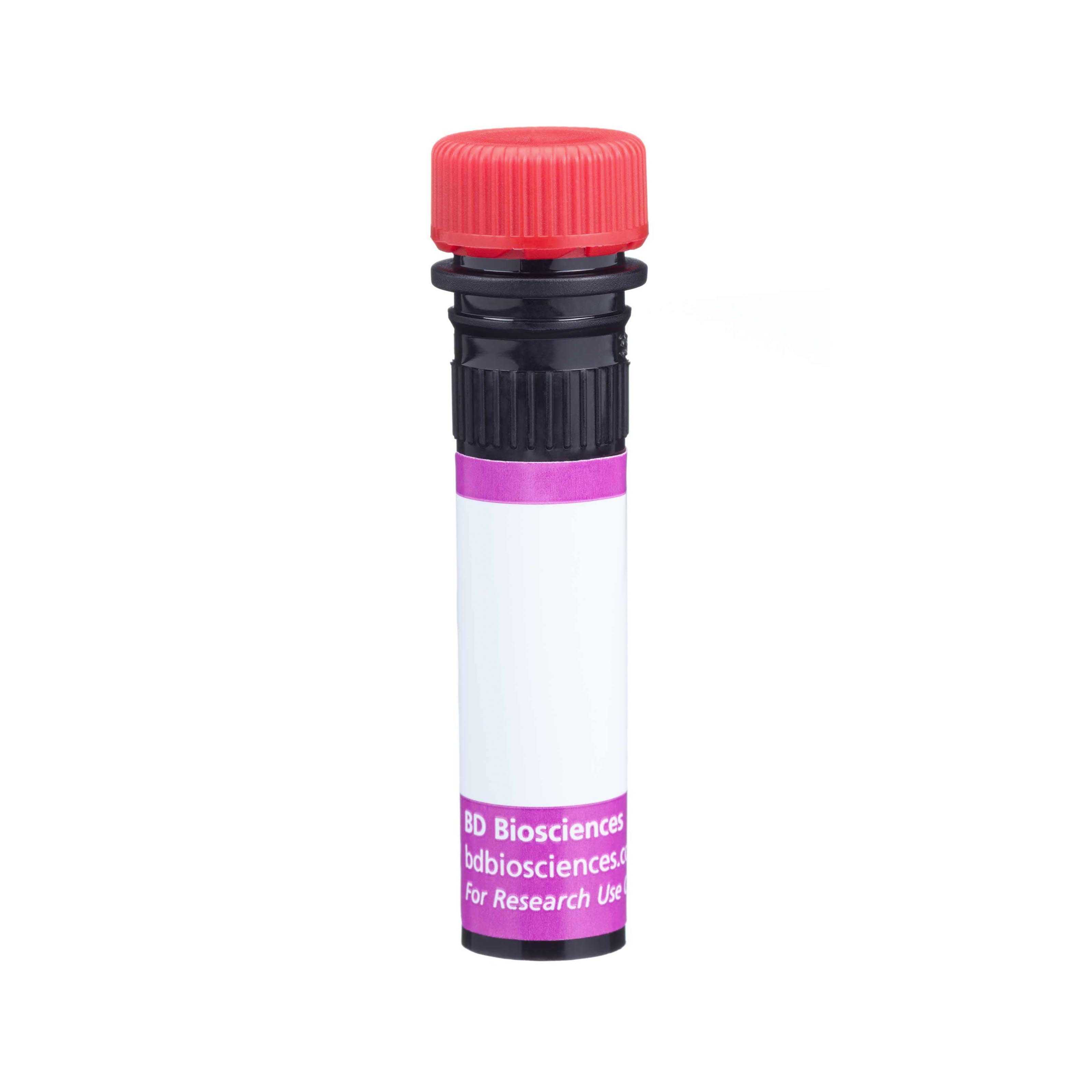
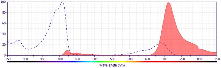
The antibody was conjugated to BD Horizon BV711 which is part of the BD Horizon Brilliant™ Violet family of dyes. This dye is a tandem fluorochrome of BD Horizon BV421 with an Ex Max of 405-nm and an acceptor dye with an Em Max at 711-nm. BD Horizon BV711 can be excited by the violet laser and detected in a filter used to detect Cy™5.5 / Alexa Fluor® 700-like dyes (eg, 712/20-nm filter). Due to the excitation and emission characteristics of the acceptor dye, there may be moderate spillover into the Alexa Fluor® 700 and PerCP-Cy5.5 detectors. However, the spillover can be corrected through compensation as with any other dye combination.
Development References (4)
-
Lanier LL, Shibuya A, Burns G. CD226 (DNAM-1, PTA1, Tlisa). In: Mason D. David Mason .. et al., ed. Leucocyte typing VII : white cell differentiation antigens : proceedings of the Seventh International Workshop and Conference held in Harrogate, United Kingdom. Oxford: Oxford University Press; 2002:921-922.
-
Shibuya A, Campbell D, Hannum C, et al. DNAM-1, a novel adhesion molecule involved in the cytolytic function of T lymphocytes. Immunity. 1996; 4(6):573-581. (Immunogen: Bioassay, Blocking, Flow cytometry, Functional assay, Immunoaffinity chromatography, Inhibition, Western blot). View Reference
-
Shibuya A, Lanier LL, Phillips JH. Protein kinase C is involved in the regulation of both signaling and adhesion mediated by DNAX accessory molecule-1 receptor. J Immunol. 1998; 161(4):1671-1676. (Clone-specific: Bioassay, Blocking, Cytotoxicity, Flow cytometry, Functional assay, Immunoprecipitation). View Reference
-
Zola H, Swart B, Boumsell L, Mason DY. Human Leucocyte Differentiation Antigen nomenclature: update on CD nomenclature. Report of IUIS/WHO Subcommittee.. J Immunol Methods. 2003; 275(1-2):1-8. (Clone-specific: Flow cytometry). View Reference
Please refer to Support Documents for Quality Certificates
Global - Refer to manufacturer's instructions for use and related User Manuals and Technical data sheets before using this products as described
Comparisons, where applicable, are made against older BD Technology, manual methods or are general performance claims. Comparisons are not made against non-BD technologies, unless otherwise noted.
For Research Use Only. Not for use in diagnostic or therapeutic procedures.
