-
Reagents
-
Flow Cytometry Reagents
- Clinical Diagnostics
-
Research Reagents
- BD Horizon RealViolet™ 828 for Flow Cytometry
- Quality and Reproducibility
- Single Color Antibodies RUO
- Panels Multicolor Cocktails RUO
- Flow Cytometry Controls and Lysates
- buffers and Supporting Reagents RUO
- Cell Function Analysis Stains Dyes
- Single Color Antibodies
- Compensation Beads
- BD Horizon™ Human T Cell Backbone Panel
- BD Pharmingen™ MonoBlock™ Leukocyte Staining Buffer
- BV605 Transition
- BD Horizon RealBlue™ 670 for Flow Cytometry
- BD Horizon RealBlue™ 780 for Flow Cytometry
- BD Horizon RealYellow™ 586
- BD Horizon RealYellow™ 610
- BD Horizon RealYellow™ 703
- BD Horizon RealBlue™ 824 for Flow Cytometry
- BD Horizon RealYellow™ 743
- BD Horizon RealYellow™ 775
- BD Horizon RealRed™ 688 Reagents
- Clinical Discovery
-
Western Blotting and Molecular Reagents
- Immunoassay Reagents
-
Single-Cell Multiomics Reagents
- BD® AbSeq Assay
-
BD® Single-Cell Multiplexing Kit
-
BD Rhapsody™ ATAC-Seq Assays
-
BD Rhapsody™ Whole Transcriptome Analysis (WTA) Amplification Kit
-
BD Rhapsody™ TCR/BCR Next Multiomic Assays
-
BD Rhapsody™ Targeted mRNA Kits
-
BD Rhapsody™ Accessory Kits
-
BD Rhapsody™ TCR/BCR Profiling Assays for Human and Mouse
- BD® OMICS-One Protein Panels
-
Functional Assays
-
Microscopy and Imaging Reagents
-
Cell Preparation and Separation Reagents
-
Flow Cytometry Reagents
-
-
-
-
- BD Horizon RealViolet™ 828 for Flow Cytometry
- Quality and Reproducibility
- Single Color Antibodies RUO
- Panels Multicolor Cocktails RUO
- Flow Cytometry Controls and Lysates
- buffers and Supporting Reagents RUO
- Cell Function Analysis Stains Dyes
- Single Color Antibodies
- Compensation Beads
- BD Horizon™ Human T Cell Backbone Panel
- BD Pharmingen™ MonoBlock™ Leukocyte Staining Buffer
- BV605 Transition
- BD Horizon RealBlue™ 670 for Flow Cytometry
- BD Horizon RealBlue™ 780 for Flow Cytometry
- BD Horizon RealYellow™ 586
- BD Horizon RealYellow™ 610
- BD Horizon RealYellow™ 703
- BD Horizon RealBlue™ 824 for Flow Cytometry
- BD Horizon RealYellow™ 743
- BD Horizon RealYellow™ 775
- BD Horizon RealRed™ 688 Reagents
-
-
-
- Brazil (English)
-
Change location/language
Old Browser
Looks like you're visiting us from {countryName}.
Would you like to stay on the current location site or be switched to your location?
BD Horizon™ BV510 Mouse Anti-Human Granzyme B
Clone GB11 (RUO)
Two-color flow cytometric analysis of Granzyme B expression by peripheral blood lymphocytes. Human peripheral blood mononuclear cells were fixed and permeabilized with BD Cytofix/Cytoperm™ Fixation and Permeabilization Solution (Cat. No. 554722). The cells were then stained with Alexa Fluor® 647 Mouse Anti-Human CD8 antibody (Cat. No. 557708) and either BD Horizon™ BV510 Mouse IgG1 Isotype Control (Cat. No. 562946; Left Panel) or BD Horizon™ BV510 Mouse Anti-Human Granzyme B antibody (Cat. No. 563388 ; Right Panel). Two-color flow cytometric dot plots showing the correlated expression patterns of Granzyme B (or Ig Isotype control staining) versus CD8 were derived from gated events with the forward and side light-scatter characteristics of intact lymphocytes. Flow cytometric analysis was performed using a BD™ LSR II Flow Cytometer System.
Regulatory Status Legend
Any use of products other than the permitted use without the express written authorization of Becton, Dickinson and Company is strictly prohibited.
Preparation And Storage
Recommended Assay Procedures
BD® CompBeads can be used as surrogates to assess fluorescence spillover (Compensation). When fluorochrome conjugated antibodies are bound to CompBeads, they have spectral properties very similar to cells. However, for some fluorochromes there can be small differences in spectral emissions compared to cells, resulting in spillover values that differ when compared to biological controls. It is strongly recommended that when using a reagent for the first time, users compare the spillover on cells and CompBead to ensure that BD® CompBeads are appropriate for your specific cellular application.
For optimal and reproducible results, BD Horizon Brilliant™ Stain Buffer should be used anytime BD Horizon Brilliant dyes are used in a multicolor flow cytometry panel. Fluorescent dye interactions may cause staining artifacts which may affect data interpretation. The BD Horizon Brilliant Stain Buffer was designed to minimize these interactions. When BD Horizon Brilliant Stain Buffer is used in the multicolor panel, it should also be used in the corresponding compensation controls for all dyes to achieve the most accurate compensation. For the most accurate compensation, compensation controls created with either cells or beads should be exposed to BD Horizon Brilliant Stain Buffer for the same length of time as the corresponding multicolor panel. More information can be found in the Technical Data Sheet of the BD Horizon Brilliant Stain Buffer (Cat. No. 563794/566349) or the BD Horizon Brilliant Stain Buffer Plus (Cat. No. 566385).
Product Notices
- Please refer to www.bdbiosciences.com/us/s/resources for technical protocols.
- Source of all serum proteins is from USDA inspected abattoirs located in the United States.
- Caution: Sodium azide yields highly toxic hydrazoic acid under acidic conditions. Dilute azide compounds in running water before discarding to avoid accumulation of potentially explosive deposits in plumbing.
- This reagent has been pre-diluted for use at the recommended Volume per Test. We typically use 1 × 10^6 cells in a 100-µl experimental sample (a test).
- An isotype control should be used at the same concentration as the antibody of interest.
- For fluorochrome spectra and suitable instrument settings, please refer to our Multicolor Flow Cytometry web page at www.bdbiosciences.com/colors.
- Species cross-reactivity detected in product development may not have been confirmed on every format and/or application.
- Human donor specific background has been observed in relation to the presence of anti-polyethylene glycol (PEG) antibodies, developed as a result of certain vaccines containing PEG, including some COVID-19 vaccines. We recommend use of BD Horizon Brilliant™ Stain Buffer in your experiments to help mitigate potential background. For more information visit https://www.bdbiosciences.com/en-us/support/product-notices.
- Please refer to http://regdocs.bd.com to access safety data sheets (SDS).
- For U.S. patents that may apply, see bd.com/patents.
Companion Products
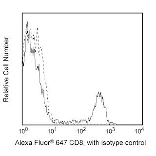
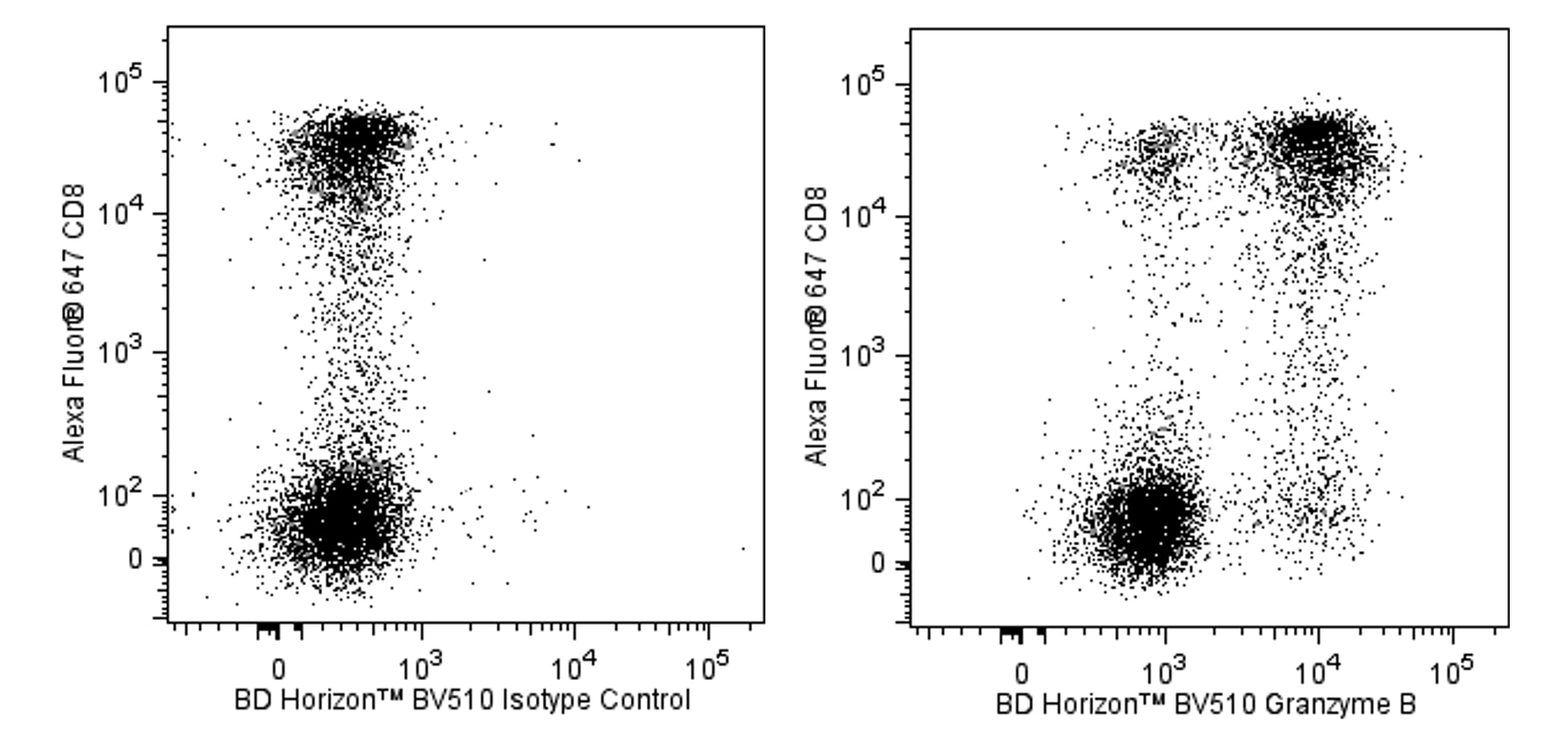
The GB11 antibody specifically reacts with human granzyme B, a serine protease of approximately 32 kDa. Granzyme B is stored in the granules of cytotoxic T lymphocytes and NK cells along with the pore-forming protein perforin. In the classic model of target cell lysis, perforins create holes in the target cell membrane allowing entrance of granzymes. Granzyme B has been shown to act on specific substrates including caspase-3, -7, -9, and -10 which in turn give rise to enzymes that mediate apoptosis. Granzyme B may also be involved in the hydrolysis of extracellular matrix components. Detectable levels of granzyme B have been detected in sera from healthy volunteers. The immunogen used to generate the GB11 hybridoma was human granzyme B isolated from an NK cell line.
Development References (8)
-
Hamann D, Baars PA, Rep MH. Phenotypic and functional separation of memory and effector human CD8+ T cells. J Exp Med. 1997; 186(9):1407-1418. (Clone-specific). View Reference
-
Poe M, Blake JT, Boulton DA. Human cytotoxic lymphocyte granzyme B. Its purification from granules and the characterization of substrate and inhibitor specificity. J Biol Chem. 1991; 266(1):98-103. (Biology). View Reference
-
Ronday HK, van der Laan WH, Tak PP et al. Human granzyme B mediates cartilage proteoglycan degradation and is expressed at the invasive front of the synovium in rheumatoid arthritis. Rheumatology (Oxford). 2001; 40:55-61. (Biology). View Reference
-
Smyth MJ, Kelly JM, Sutton VR et al. Unlocking the secrets of cytotoxic granule proteins. J Leukoc Biol. 2001; 70:18-29. (Biology). View Reference
-
Spaeny-Dekking EH, Hanna WL, Wolbink AM et al. Extracellular granzymes A and B in humans: detection of native species during CTL responses in vitro and in vivo. J Immunol. 1998; 160:3610. (Immunogen: ELISA, Radioimmunoassay). View Reference
-
Trapani JA, Klein JL, White PC, and Dupont B. Molecular cloning of an inducible serine esterase gene from human cytotoxic lymphocytes. Proc Natl Acad Sci U S A. 1988; 5:6924-6928. (Biology). View Reference
-
Trapani JA, Smyth MJ, Apostolidis VA, Dawson M, and Browne KA. Granule serine proteases are normal nuclear constituents of natural killer cells. J Biol Chem. 1994; 269:18359-18365. (Biology). View Reference
-
Wever PC, Van Der Vliet HJ, Spaeny LH . The CD8+ granzyme B+ T-cell subset in peripheral blood from healthy individuals contains activated and apoptosis-prone cells. Immunology. 1998; 93(3):383-389. (Immunogen: ELISA, Flow cytometry, Immunocytochemistry (cytospins), Immunoprecipitation, Radioimmunoassay). View Reference
Please refer to Support Documents for Quality Certificates
Global - Refer to manufacturer's instructions for use and related User Manuals and Technical data sheets before using this products as described
Comparisons, where applicable, are made against older BD Technology, manual methods or are general performance claims. Comparisons are not made against non-BD technologies, unless otherwise noted.
For Research Use Only. Not for use in diagnostic or therapeutic procedures.