







Preparation And Storage


Product Notices
- Since applications vary, each investigator should titrate the reagent to obtain optimal results.
- An isotype control should be used at the same concentration as the antibody of interest.
- Caution: Sodium azide yields highly toxic hydrazoic acid under acidic conditions. Dilute azide compounds in running water before discarding to avoid accumulation of potentially explosive deposits in plumbing.
- Source of all serum proteins is from USDA inspected abattoirs located in the United States.
- For fluorochrome spectra and suitable instrument settings, please refer to our Multicolor Flow Cytometry web page at www.bdbiosciences.com/colors.
- Please refer to www.bdbiosciences.com/us/s/resources for technical protocols.
Companion Products
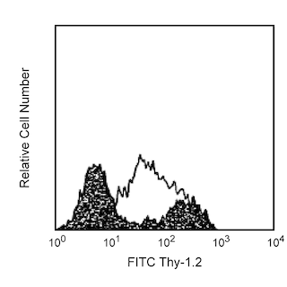



The GL1 antibody specifically recognizes the B7-2 (CD86) costimulatory molecule expressed on a broad spectrum of leukocytes, including B lymphocytes, T lymphocytes, thioglycollate-induced peritoneal macrophages, dendritic cells and astrocytes. CD86 is expressed at low levels by freshly explanted peripheral B and T cells, and its expression is substantially increased by a variety of T cell- and B cell-specific stimuli with a peak expression after 18-42 hours of culture. In contrast to most naive CD4+ T cells, memory CD4+ T cells express B7-2, both at the mRNA and protein level. CD86, a ligand for CD28 and CD152 (CTLA-4), is one of the accessory molecules that plays an important role in T cell-B cell costimulatory interactions. It has been shown to be involved in immunoglobulin class-switching and triggering of mouse NK cell-mediated cytotoxicity. CD80 (B7-1) is an alternate ligand for CD28 and CD152 (CTLA-4). GL1 antibody reportedly blocks MLR and stimulation of T cells by natural antigen-presenting cells. In addition, a mixture of anti-B7-1 and anti B7-2 (GL1) mAbs reportedly inhibits the in vitro interaction of CTLA-4 with its ligand and the in vivo priming of cytotoxic T lymphocytes.
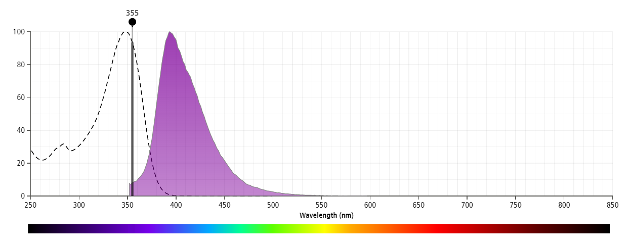
The antibody was conjugated to BD Horizon™ BUV395 which is part of the BD Horizon Brilliant™ Ultraviolet family of dyes. This dye is optimal for multicolor flow cytometry because it has little to no spillover into other detectors. With an Ex Max at 348 nm and an Em Max at 395 nm, BD Horizon BUV395 can be excited with a 355 nm laser and detected with a 379/28 filter.
Development References (14)
-
Bluestone JA. New perspectives of CD28-B7-mediated T cell costimulation. Immunity. 1995; 2(6):555-559. (Biology). View Reference
-
Borriello F, Sethna MP, Boyd SD, et al. B7-1 and B7-2 have overlapping, critical roles in immunoglobulin class switching and germinal center formation. Immunity. 1997; 6(3):303-313. (Biology). View Reference
-
Freeman GJ, Borriello F, Hodes RJ, et al. Uncovering of functional alternative CTLA-4 counter-receptor in B7-deficient mice. Science. 1993; 262(5135):907-909. (Biology). View Reference
-
Hakamada-Taguchi R, Kato T, Ushijima H, Murakami M, Uede T, Nariuchi H. Expression and co-stimulatory function of B7-2 on murine CD4+ T cells. Eur J Immunol. 1998; 28(3):865-873. (Biology). View Reference
-
Hathcock KS, Laszlo G, Dickler HB, Bradshaw J, Linsley P, Hodes RJ. Identification of an alternative CTLA-4 ligand costimulatory for T cell activation. Science. 1993; 262(5135):905-907. (Immunogen: Blocking, Immunoprecipitation). View Reference
-
Hathcock KS, Laszlo G, Pucillo C, Linsley P, Hodes RJ. Comparative analysis of B7-1 and B7-2 costimulatory ligands: expression and function. J Exp Med. 1994; 180(2):631-640. (Clone-specific: Flow cytometry, Inhibition). View Reference
-
Inaba K, Witmer-Pack M, Inaba M, et al. The tissue distribution of the B7-2 costimulator in mice: abundant expression on dendritic cells in situ and during maturation in vitro. J Exp Med. 1994; 180(5):1849-1860. (Clone-specific: Functional assay, Immunohistochemistry, Inhibition). View Reference
-
Krummel MF, Allison JP. CD28 and CTLA-4 have opposing effects on the response of T cells to stimulation. J Exp Med. 1995; 182(2):459-465. (Clone-specific: Blocking). View Reference
-
Larsen CP, Ritchie SC, Hendrix R, et al. Regulation of immunostimulatory function and costimulatory molecule (B7-1 and B7-2) expression on murine dendritic cells. J Immunol. 1994; 152(11):5208-5219. (Clone-specific: Flow cytometry). View Reference
-
Lenschow DJ, Su GH, Zuckerman LA, et al. Expression and functional significance of an additional ligand for CTLA-4. Proc Natl Acad Sci U S A. 1993; 90(23):11054-11058. (Biology). View Reference
-
Liu Y, Wenger RH, Zhao M, Nielsen PJ. Distinct costimulatory molecules are required for the induction of effector and memory cytotoxic T lymphocytes. J Exp Med. 1997; 185(2):251-262. (Clone-specific: Blocking). View Reference
-
Martin-Fontecha A, Assarsson E, Carbone E, Karre K, Ljunggren HG. Triggering of murine NK cells by CD40 and CD86 (B7-2). J Immunol. 1999; 162(10):5910-5916. (Biology). View Reference
-
Turley SJ, Inaba K, Garrett WS, et al. Transport of peptide-MHC class II complexes in developing dendritic cells. Science. 2000; 288(5465):522-527. (Clone-specific: Electron microscopy, Fluorescence microscopy). View Reference
-
Yang G, Mizuno MT, Hellstrom KE, Chen L. B7-negative versus B7-positive P815 tumor: differential requirements for priming of an antitumor immune response in lymph nodes. J Immunol. 1997; 158(2):851-858. (Clone-specific: Immunohistochemistry). View Reference
Please refer to Support Documents for Quality Certificates
Global - Refer to manufacturer's instructions for use and related User Manuals and Technical data sheets before using this products as described
Comparisons, where applicable, are made against older BD Technology, manual methods or are general performance claims. Comparisons are not made against non-BD technologies, unless otherwise noted.
For Research Use Only. Not for use in diagnostic or therapeutic procedures.
