Old Browser
Looks like you're visiting us from {countryName}.
Would you like to stay on the current country site or be switched to your country?
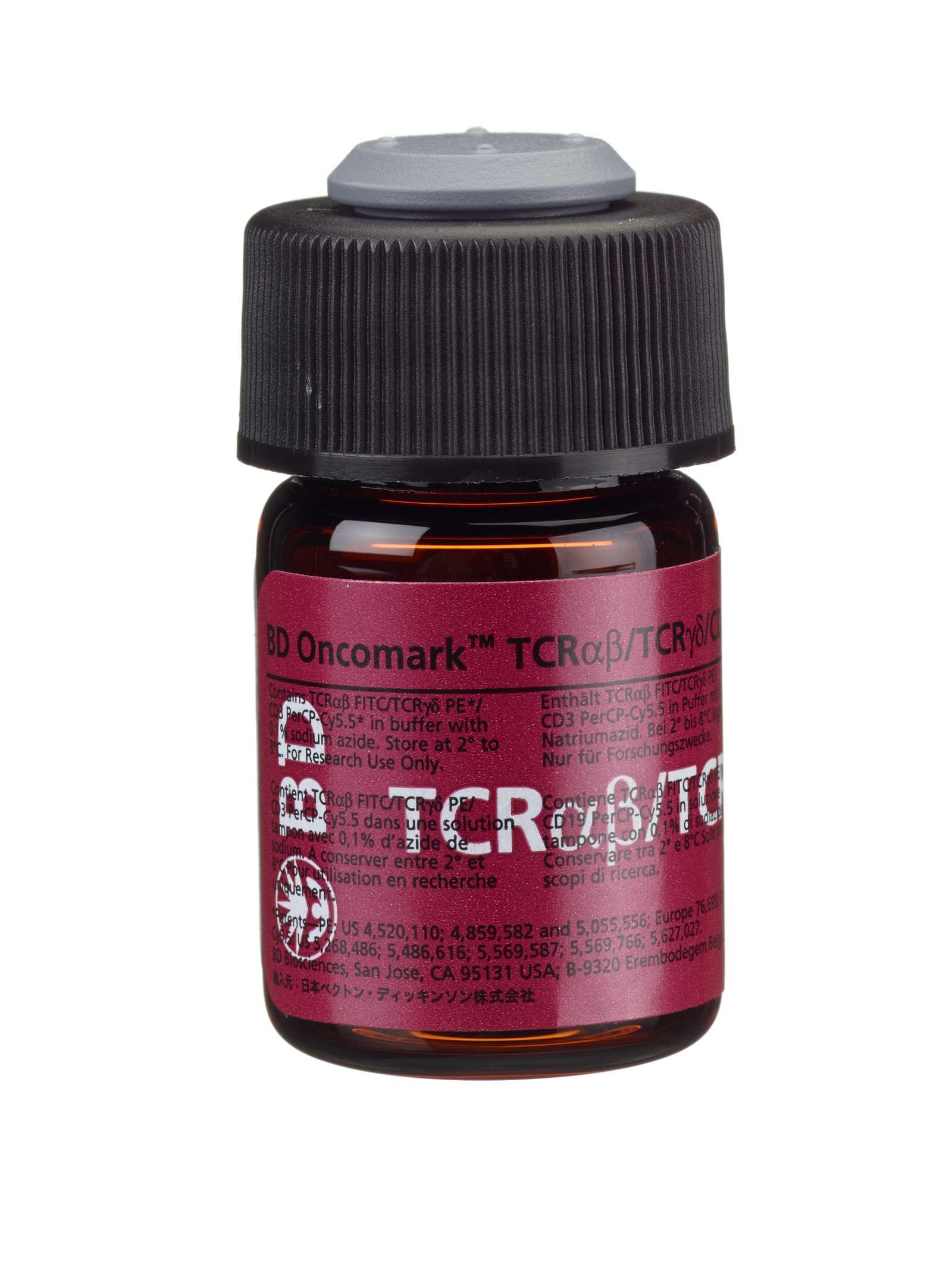
BD Oncomark™ Anti-Human TCR-αβ FITC/TCR-γδ PE/CD3 PerCP-Cy™5.5
(RUO (GMP))

Anti-Human TCR-αβ FITC/TCR-γδ PE/CD3 PerCP-Cy™5.5
Regulatory Status Legend
Any use of products other than the permitted use without the express written authorization of Becton, Dickinson and Company is strictly prohibited.
Description
Anti–TCR-α/β-1, clone WT31, is derived from hybridization of mouse Sp2/0-Ag14 myeloma cells with spleen cells from BALB/c mice immunized with human thymocytes. Anti–TCR-γ/δ-1, clone 11F2, is derived from hybridization of mouse Sp2/0 myeloma cells with spleen cells from BALB/c mice immunized with a Sepharose® bead/CD3/γ/δ TCR complex. CD3, clone SK7, is derived from hybridization of mouse NS-1 myeloma cells with spleen cells from BALB/c mice immunized with human thymocytes. Anti–TCR-α/β-1 recognizes a conformational epitope formed by the T-cell receptor (TCR) for antigen and the CD3 epsilon chain. The α/β TCR is a disulfide-linked 80-kilodalton (kd) heterodimer consisting of a 44-kd α chain and a 37-kd β chain. Anti–TCR-γ/δ-1 reacts with a framework epitope of the γ/δ T-cell antigen receptor (TCR). The γ/δ TCR is a heterodimeric glycoprotein that is noncovalently associated with the CD3 antigen. The γ and δ TCR chains are composed of constant and variable regions, each encoded by distinct gene segments. The γ chain forms either disulfide-linked or non–disulfide-linked heterodimers with the δ subunit. CD3 reacts with the epsilon chain of the CD3 antigen/T-cell antigen receptor (TCR) complex. This complex is composed of at least six proteins that range in molecular weight from 20 to 30 kd. The antigen recognized by CD3 antibodies is noncovalently associated with either α/β or γ/δ TCR (70 to 90 kd).
Preparation And Storage
Store vials at 2° to 8°C. Do not freeze reagents; protect them from prolonged exposure to light. Each reagent is stable for the period shown on the bottle label when stored as directed.
| Description | Clone | Isotype | EntrezGene ID |
|---|---|---|---|
| gamma delta TCR PE | 11F2 | IgG1, | N/A |
| CD3 PerCP-Cy5.5 | SK7 | IgG1, κ | N/A |
| TCR αβ FITC | WT31 | IgG1, κ | N/A |
Development References (20)
-
Bolhuis RLH, Beiske K, Klepper LK, Alfsen C. Knapp W, Dörken B, Gilks WR, et al, ed. Leucocyte Typing IV: White Cell Differentiation Antigens. New York, NY: Oxford University Press; 1989:302.
-
Borst J, van Dongen JJ, Bolhuis RL, et al. Distinct molecular forms of human T cell receptor gamma/delta detected on viable T cells by a monoclonal antibody.. J Exp Med. 1988; 167(5):1625-44. (Biology). View Reference
-
Brenner M, Groh V, Porcelli S, et al. Knapp W, Dörken B, Gilks W, et al, ed. Leucocyte Typing IV: White Cell Differentiation Antigens. New York: Oxford University Press; 1989:1049-1053.
-
Brenner MB, Groh V, Porcelli SA, et al. Knapp W, Dörken B, Gilks WR, et al, ed. Leucocyte Typing IV: White Cell Differentiation Antigens. 1986:145-149.
-
Campana D, Coustan-Smith E, Janossy G. Knapp W, Dörken B, Gilks WR, et al, ed. Leucocyte Typing IV: White Cell Differentiation Antigens. New York, NY: Oxford University Press; 1989:297-298.
-
Centers for Disease Control. Update: universal precautions for prevention of transmission of human immunodeficiency virus, hepatitis B virus, and other bloodborne pathogens in healthcare settings. MMWR. 1988; 37:377-388. (Biology).
-
Clevers H, Alarcón B, Wileman T, Terhorst C. The T cell receptor/CD3 complex: a dynamic protein ensemble. Annual Rev Immunol. 1988; 6:629. (Biology).
-
Falini B, Flenghi L, Fagioli M, et al. Knapp W, Dörken B, Gilks WR, et al, ed. Leucocyte Typing IV: White Cell Differentiation Antigens. New York, NY: Oxford University Press; 1989:303-305.
-
Lanier LL, Ruitenberg J, Bolhuis RL, Borst J, Phillips JH, Testi R. Structural and serological heterogeneity of gamma/delta T cell antigen receptor expression in thymus and peripheral blood.. Eur J Immunol. 1988; 18(12):1985-92. (Biology). View Reference
-
Lanier LL, Serafini AT, Ruitenberg JJ, et al. The γ T-cell antigen receptor. J Clin Immunol. 1987; 7:429-440. (Biology).
-
Lanier LL, Weiss A. Presence of Ti (WT31) negative T lymphocytes in peripheral blood and thymus. Nature. 1986; 324:268. (Biology).
-
Ledbetter JA, Uckun FM. Knapp W, Dörken B, Gilks WR, et al, ed. Leucocyte Typing IV: White Cell Differentiation Antigens. New York, NY: Oxford University Press; 1989:312-314.
-
Matsuo Y, Ariyasu T, Brenner MB, Imanishi J, Yokoyama MM, Minowada J. Knapp W, Dörken B, Gilks WR, et al, ed. Leucocyte Typing IV: White Cell Differentiation Antigens. New York, NY: Oxford University Press; 1989:299-301.
-
Oettgen HC, Kappler J, Tax WJ, Terhorst C. Characterization of the two heavy chains of the T3 complex on the surface of human T lymphocytes. J Biol Chem. 1984; 259(19):12039-12048. (Biology). View Reference
-
Reichert T, DeBruyere M, Deneys V, et al. Lymphocyte subset reference ranges in adult Caucasians. Clin Immunol Immunopathol. 1991; 60(2):190-208. (Biology). View Reference
-
Slameron A, Sanchez-Madrid F, Ursa MA, Fresno M, and Alarcon B. A conformational epitope expressed upon association of the CD3-epsilon with either CD3-delta or CD3-gamma is the main target for recognition by anti-CD3 monoclonal antibodies. J Immunol. 1991; 147:3047-3052. (Biology).
-
Spits H, Borst J, Tax W, Capel PJ, Terhorst C, de Vries JE. Characteristics of a monoclonal antibody (WT-31) that recognizes a common epitope on the human T cell receptor for antigen.. J Immunol. 1985; 135(3):1922-8. (Biology). View Reference
-
Testi R, Lanier LL. Functional expression of CD28 on T cell antigen receptor γ/δ-bearing T lymphocytes. Eur J Immunol. 1989; 19:185-188. (Biology).
-
Weiss A, Newton M, Crommie D. Expression of T3 in association with a molecule distinct from the T-cell antigen receptor heterodimer.. Proc Natl Acad Sci USA. 1986; 83(18):6998-7002. (Biology). View Reference
-
van Dongen JJM, Krissansen GW, Wolvers-Tettero ILM, et al. Cytoplasmic expression of the CD3 antigen as a diagnostic marker for immature T-cell malignancies. Blood. 1988; 71:603-612. (Biology).
Please refer to Support Documents for Quality Certificates
Global - Refer to manufacturer's instructions for use and related User Manuals and Technical data sheets before using this products as described
Comparisons, where applicable, are made against older BD Technology, manual methods or are general performance claims. Comparisons are not made against non-BD technologies, unless otherwise noted.
For Research Use Only. Not for use in diagnostic or therapeutic procedures.
Although not required, these products are manufactured in accordance with Good Manufacturing Practices.
Report a Site Issue
This form is intended to help us improve our website experience. For other support, please visit our Contact Us page.