-
Reagents
- Flow Cytometry Reagents
-
Western Blotting and Molecular Reagents
- Immunoassay Reagents
-
Single-Cell Multiomics Reagents
- BD® OMICS-Guard Sample Preservation Buffer
- BD® AbSeq Assay
- BD® Single-Cell Multiplexing Kit
- BD Rhapsody™ ATAC-Seq Assays
- BD Rhapsody™ Whole Transcriptome Analysis (WTA) Amplification Kit
- BD Rhapsody™ TCR/BCR Next Multiomic Assays
- BD Rhapsody™ Targeted mRNA Kits
- BD Rhapsody™ Accessory Kits
- BD® OMICS-One Protein Panels
-
Functional Assays
-
Microscopy and Imaging Reagents
-
Cell Preparation and Separation Reagents
-
- BD® OMICS-Guard Sample Preservation Buffer
- BD® AbSeq Assay
- BD® Single-Cell Multiplexing Kit
- BD Rhapsody™ ATAC-Seq Assays
- BD Rhapsody™ Whole Transcriptome Analysis (WTA) Amplification Kit
- BD Rhapsody™ TCR/BCR Next Multiomic Assays
- BD Rhapsody™ Targeted mRNA Kits
- BD Rhapsody™ Accessory Kits
- BD® OMICS-One Protein Panels
- Australia (English)
-
Change country/language
Old Browser
Looks like you're visiting us from United States.
Would you like to stay on the current country site or be switched to your country?
BD Transduction Laboratories™ Purified Mouse Anti-Cadherin-5
Clone 75/Cadherin-5 (RUO)


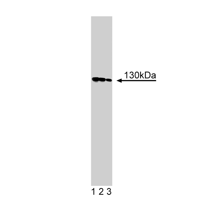
Western blot analysis of Cadherin-5 on human endothelial cell lysate. Lane 1: 1:250, lane 2: 1:500, lane 3: 1:1000 dilution of anti-Cadherin-5 antibody.

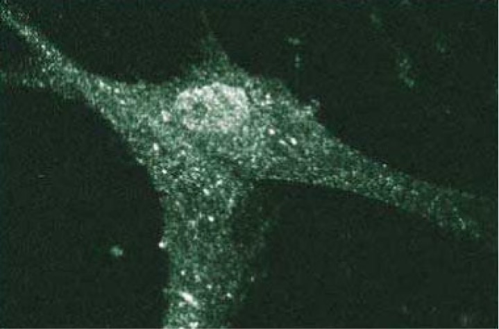
Immunofluorescent staining of Human Fibroblasts with anti-Cadherin-5 antibody.


BD Transduction Laboratories™ Purified Mouse Anti-Cadherin-5

BD Transduction Laboratories™ Purified Mouse Anti-Cadherin-5

Regulatory Status Legend
Any use of products other than the permitted use without the express written authorization of Becton, Dickinson and Company is strictly prohibited.
Preparation And Storage
Recommended Assay Procedures
Western blot: Please refer to http://www.bdbiosciences.com/pharmingen/protocols/Western_Blotting.shtml.
Product Notices
- Since applications vary, each investigator should titrate the reagent to obtain optimal results.
- Please refer to www.bdbiosciences.com/us/s/resources for technical protocols.
- Caution: Sodium azide yields highly toxic hydrazoic acid under acidic conditions. Dilute azide compounds in running water before discarding to avoid accumulation of potentially explosive deposits in plumbing.
- Source of all serum proteins is from USDA inspected abattoirs located in the United States.
Cadherins are a family of transmembrane glycoproteins involved in the Ca2+- dependent cell-cell adhesion that occurs in many tissues. These proteins are similar in their domain structure (45-74% amino acid conservation), Ca2+ and protease-sensitivity, and molecular weight. Cadherin-5 (VE-Cadherin or CD144) is one of a number of cadherins (cadherin-4 through -11) whose cDNAs were isolated from rat brain and retina. These cadherins have a cytoplasmic domain that is highly conserved relative to previously identified cadherins, indicating that this domain is essential for cell adhesion activity. This function is mediated by cadherin interaction with cytoskeletal proteins. However, Cadherin-5's cytoplasmic domain has the lowest degree of homology with the other cadherins. Cadherin-5 is expressed in brain and various other tissues, including umbilical cord vein endothelial cells. A new type of adhering junction has been identified in certain vascular endothelial cells. These junctions are known as "complexus adherens" and are morphologically and compositionally distinct from desmosomes and zonula adherens junctions. The complexus adherens of endothelial cells lack desmosomal cadherins as well as E-Cadherin. However, these cells are rich in Cadherin-5 which colocalizes with desmoplakin and γ-Catenin (plakoglobin).
Development References (5)
-
Corada M, Liao F, Lindgren M. Monoclonal antibodies directed to different regions of vascular endothelial cadherin extracellular domain affect adhesion and clustering of the protein and modulate endothelial permeability. Blood. 2001; 97(6):1679-1684. (Clone-specific: Immunofluorescence, Western blot). View Reference
-
Corada M, Zanetta L, Orsenigo F. A monoclonal antibody to vascular endothelial-cadherin inhibits tumor angiogenesis without side effects on endothelial permeability. Blood. 2002; 100(3):905-911. (Clone-specific: Flow cytometry, Western blot). View Reference
-
Rahimi N, Kazlauskas A. A role for cadherin-5 in regulation of vascular endothelial growth factor receptor 2 activity in endothelial cells. Mol Biol Cell. 1999; 10:3401-3407. (Clone-specific: Functional assay). View Reference
-
Schmelz M, Franke WW. Complexus adhaerentes, a new group of desmoplakin-containing junctions in endothelial cells: the syndesmos connecting retothelial cells of lymph nodes. J Cell Biol. 1993; 61(2):274-289. (Biology). View Reference
-
Suzuki S, Sano K, Tanihara H. Diversity of the cadherin family: evidence for eight new cadherins in nervous tissue. Cell Regul. 1991; 2(4):261-270. (Biology). View Reference
Please refer to Support Documents for Quality Certificates
Global - Refer to manufacturer's instructions for use and related User Manuals and Technical data sheets before using this products as described
Comparisons, where applicable, are made against older BD Technology, manual methods or are general performance claims. Comparisons are not made against non-BD technologies, unless otherwise noted.
For Research Use Only. Not for use in diagnostic or therapeutic procedures.
Refer to manufacturer's instructions for use and related User Manuals and Technical Data Sheets before using this product as described.
Comparisons, where applicable, are made against older BD technology, manual methods or are general performance claims. Comparisons are not made against non-BD technologies, unless otherwise noted.