








Preparation And Storage


Recommended Assay Procedures
Functional Studies: N-CAM is expressed on many cell and tissue types including neural tissue, developing and diseased smooth and cardiac muscle, NK lymphocytes, and a small percentage of T lymphocytes. Anti-N-CAM-16 monoclonal antibody (clone NCAM16.2) can be used with other anti-cell adhesion molecule monoclonal antibodies to better understand the role of N-CAM in nervous system inflammation and tumor cell metastasis. This antibody has been used with monoclonal antibodies to integrins αL, α4, β1, and β2, ICAM-1, VCAM-1 and E-selectin, and other monoclonal antibodies to N-CAM to study mononuclear leukocyte adhesion to neuroblastoma and cortical neural cells. NK lymphocytes and the subset of T lymphocytes that express N-CAM are unique in their ability to mediate direct, non-major histocompatibility complex-restricted cytotoxicity against certain tumor cells.
Immunoprecipitation: Anti-N-CAM-16 monoclonal antibody (clone NCAM16.2) immunoprecipitates N-CAM from human brain, KG-1a cells, leukemia cells, and N-CAM-expressing neuroblastoma cells. Immunohistology: Anti-N-CAM-16 monoclonal antibody (clone NCAM16.2) can be used for staining of frozen or paraffin-embedded sections of human or rat neural and skeletal muscle tissues by indirect immunofluoresscence or immunoperoxidase metods, or staining of cultured LAN-1 human neuroblastoma cells by indirect immunofluorescence.
Enzyme-linked immunosorbent assay (ELISA)/Radioimmunoassay (RIA): Anti-N-CAM-16 monoclonal antibody (clone NCAM16.2) can be used in an ELISA to detect N-CAM in brain extracts.
Product Notices
- Since applications vary, each investigator should titrate the reagent to obtain optimal results.
- Please refer to www.bdbiosciences.com/us/s/resources for technical protocols.
- Caution: Sodium azide yields highly toxic hydrazoic acid under acidic conditions. Dilute azide compounds in running water before discarding to avoid accumulation of potentially explosive deposits in plumbing.
Companion Products
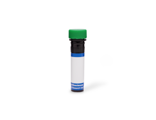

The NCAM16.2 monoclonal antibody specifically binds to human CD56. It recognizes an extracellular immunoglobulin-like domain common to 120, 140, and 180 kDa forms of CD56, also known as the neural cell adhesion molecule (NCAM), NKH1 or MSK39. The CD56 antigen is expressed on approximately 10% to 25% of peripheral blood lymphocytes. It is present on essentially all resting and activated CD16+ natural killer (NK) lymphocytes and approximately 5% of CD3+ peripheral blood lymphocytes. CD3+ CD56+ T lymphocytes comprise a unique subset of cytotoxic T lymphocytes that mediates non-major histocompatibility complex (MHC)-restricted cytotoxicity. CD56 antigen density on NK lymphocytes increases upon cellular activation. The CD56 antigen is involved in neuronal homotypic cell adhesion and cell differentiation during embryogenesis. CD16+ CD56+ NK cells demonstrate reciprocal transfer of an activation state with dendritic cells.
Development References (12)
-
Birdsall HH, Lane C, Ramser MN, Anderson DC. Induction of VCAM-1 and ICAM-1 on human neural cells and mechanisms of mononuclear leukocyte adherence. J Immunol. 1992; 148(9):2717-2723. (Biology). View Reference
-
Cunningham BA, Hemperly JJ, Murray BA, Prediger EA, Brackenbury R, Edelman GM. Neural cell adhesion molecule: structure, immunoglobulin-like domains, cell surface modulation, and alternative RNA splicing. Science. 1987; 236(4803):799-806. (Biology). View Reference
-
Doherty P, Ashton SV, Moore SE, Walsh FS. Morphoregulatory activities of NCAM and N-cadherin can be accounted for by G protein-dependent activation of L- and N-type neuronal Ca2+ channels. Cell. 1991; 67(1):21-33. (Biology). View Reference
-
Edelman GM. Cell adhesion molecules in the regulation of animal form and tissue pattern. Annu Rev Cell Biol. 1986; 2:81-116. (Biology). View Reference
-
Hemperly JJ, Murray BA, Edelman GM, Cunningham BA. Sequence of a cDNA clone encoding the polysialic acid-rich and cytoplasmic domains of the neural cell adhesion molecule N-CAM. Proc Natl Acad Sci U S A. 1986; 83(9):3037-3041. (Biology). View Reference
-
Jin L, Hemperly JJ, Lloyd RV. Expression of neural cell adhesion molecule in normal and neoplastic human neuroendocrine tissues. Am J Pathol. 1991; 138(4):961-969. (Biology). View Reference
-
Lanier LL, Chang C, Azuma M, Ruitenberg JJ, Hemperly JJ, Phillips JH. Molecular and functional analysis of human natural killer cell-associated neural cell adhesion molecule (N-CAM/CD56). J Immunol. 1991; 146(12):4421-4426. (Biology). View Reference
-
Lanier LL, Le AM, Civin CI, Loken MR, Phillips JH. The relationship of CD16 (Leu-11) and Leu-19 (NKH-1) antigen expression on human peripheral blood NK cells and cytotoxic T lymphocytes. J Immunol. 1986; 136(12):4480-4486. (Biology). View Reference
-
Lanier LL, Le AM, Ding A. Expression of Leu-19 (NKH-1) antigen on IL 2-dependent cytotoxic and non-cytotoxic T cell lines. J Immunol. 1987; 138(7):2019-2023. (Biology). View Reference
-
Lanier LL, Testi R, Bindl J, Phillips JH. Identity of Leu-19 (CD56) leukocyte differentiation antigen and neural cell adhesion molecule. J Exp Med. 1989; 169(6):2233-2238. (Biology). View Reference
-
Schubert W, Zimmermann K, Cramer M, Starzinski-Powitz A. Lymphocyte antigen Leu-19 as a molecular marker of regeneration in human skeletal muscle. Proc Natl Acad Sci U S A. 1989; 86(1):307-311. (Biology). View Reference
-
Zocchi MR, Vidal M, Poggi A. Involvement of CD56/N-CAM molecule in the adhesion of human solid tumor cell lines to endothelial cells. Exp Cell Res. 1993; 204(1):130-135. (Biology). View Reference
Please refer to Support Documents for Quality Certificates
Global - Refer to manufacturer's instructions for use and related User Manuals and Technical data sheets before using this products as described
Comparisons, where applicable, are made against older BD Technology, manual methods or are general performance claims. Comparisons are not made against non-BD technologies, unless otherwise noted.
For Research Use Only. Not for use in diagnostic or therapeutic procedures.
Refer to manufacturer's instructions for use and related User Manuals and Technical Data Sheets before using this product as described.
Comparisons, where applicable, are made against older BD technology, manual methods or are general performance claims. Comparisons are not made against non-BD technologies, unless otherwise noted.