BD Pharmingen™ Alexa Fluor® 555 Mouse anti-BrdU
Clone 3D4 (RUO)
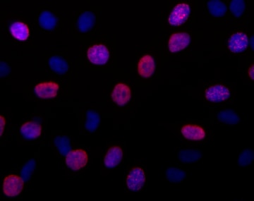
Immunofluorescent staining of HeLa cells. Cells were seeded in a BD Falcon™ 96-well Imaging Plate (Cat. No. 353219) at ~ 10,000 cells per well. After overnight culture, the cells were loaded with 20 μM BrdU for 1 hour at 37°C. After treatment, the cells were fixed, permeabilized, DNAse treated, stained with Alexa Fluor® 555 Mouse anti-BrdU (pseudo colored red), and counter stained with Hoechst 33342 (pseudo colored blue) according to the Recommended Assay Procedure. The images were captured on a BD Pathway™ 435 confocal imager with a 20x objective and merged using BD AttoVision™ software.
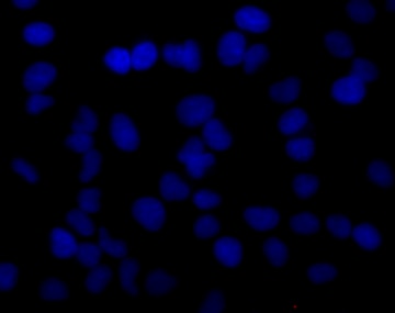
Negative control for immunofluorescent staining of HeLa cells. Cells were cultured as in the left image, but without BrdU loading. They were fixed, permeabilized, DNAse treated, stained with Alexa Fluor® 555 Mouse anti-BrdU (pseudo colored red), and counter stained with Hoechst 33342 (pseudo colored blue) according to the Recommended Assay Procedure. The images were captured on a BD Pathway™ 435 confocal imager with a 20x objective and merged using BD AttoVision™ software.
Immunofluorescent staining of HeLa cells. Cells were seeded in a BD Falcon™ 96-well Imaging Plate (Cat. No. 353219) at ~ 10,000 cells per well. After overnight culture, the cells were loaded with 20 μM BrdU for 1 hour at 37°C. After treatment, the cells were fixed, permeabilized, DNAse treated, stained with Alexa Fluor® 555 Mouse anti-BrdU (pseudo colored red), and counter stained with Hoechst 33342 (pseudo colored blue) according to the Recommended Assay Procedure. The images were captured on a BD Pathway™ 435 confocal imager with a 20x objective and merged using BD AttoVision™ software.
Negative control for immunofluorescent staining of HeLa cells. Cells were cultured as in the left image, but without BrdU loading. They were fixed, permeabilized, DNAse treated, stained with Alexa Fluor® 555 Mouse anti-BrdU (pseudo colored red), and counter stained with Hoechst 33342 (pseudo colored blue) according to the Recommended Assay Procedure. The images were captured on a BD Pathway™ 435 confocal imager with a 20x objective and merged using BD AttoVision™ software.
Preparation And Storage
Recommended Assay Procedures
Cells are pulse labeled with BrdU, fixed to maintain BrdU loading, and permeabilized for intranuclear staining of the incorporated BrdU. DNase I is used to randomly cleave the DNA, allowing the anti-BrdU monoclonal antibody to bind to the incorporated BrdU.
Materials
• Adherent cell culture growing in a BD Falcon™ 96-well Imaging Plate (Cat. No. 353219)
• 37°C incubator
• BrdU (Cat. No. 550891) diluted to 10-100 μM in tissue culture medium, prepare enough to use at 50 μl per well (see step 1 of procedure)
• BD Cytofix™ fixation buffer (Cat. No. 554655), warmed to 37°C
• BD™ Phosflow Perm Buffer III (Cat. No. 558050), at -20°C
• Phosphate-buffered saline solution (PBS), at room temperature
• BD Pharmingen™ Stain Buffer (FBS) (Cat. No. 554656), at 4°C
• 300 μg/ml sterile solution of DNase I (preferred, Sigma-Aldrich Cat. No. D4513) in PBS, prepare enough to use at 50 μl per well
• Alexa Fluor® 555 Mouse anti-BrdU monoclonal antibody diluted 1:10 in BD Pharmingen™ Stain Buffer (FBS), prepare enough to use at 50 μl per well. If additional antibodies are to be used, they should be added so that all antibodies are included in the 50 μl per well.
• 2 μg/ml solution of Hoechst 33342 (eg, Sigma-Aldrich Cat. No. B2261) in PBS, prepare enough to use at 100 μl per well
• Fluorescence imaging instrument capable of exciting Alexa Fluor® 555 and Hoechst 33342, such as the BD Pathway™ Bioimaging System
Procedure
1. Pulse the adherent cell cultures by adding 50 μl of the diluted BrdU to each well and incubating for 1-2 hrs at 37°C. Omit the BrdU from the wells that will be used as negative controls for the BrdU staining. In all subsequent steps, the BrdU-loaded and control cells should be processed in parallel.
2. Remove the medium from the wells, and fix the cells by adding 100 μl of the pre-warmed fixation buffer to each well and incubating for 10 - 20 minutes at room temperature.
3. Remove the fixative from the wells, and wash the wells twice with 100 μl of PBS.
4. Remove the PBS, and permeabilize the cells by adding 100 μl of the ice-cold BD™ Phosflow Perm Buffer III to each well and incubating for 5 - 10 minutes at room temperature.
5. Remove the Perm Buffer III from the wells, and wash the wells twice with 100 μl of PBS.
6. OPTIONAL: Remove the PBS, and block the cells by adding 100 μl of the BD Pharmingen™ Stain Buffer (FBS) to each well and incubating for 15 - 30 minutes at room temperature.
7. Remove the BD Pharmingen™ Stain Buffer (FBS), and denature the cells' DNA by adding 50 μl of the sterile DNase I solution to each well and incubating for 1 hour at 37°C.
8. Remove the DNase I solution from the wells, and wash the wells once with 100 μl of PBS.
9. Remove the PBS, and stain the cells by adding 50 μl of the diluted antibody solution to each well and incubating for 1 hour at room temperature.
10. Remove the antibody solution, and wash the wells twice with 100 μl of PBS.
11. Remove the PBS, and counter-stain the nuclei by adding 100 μl of the Hoechst 33342 solution to each well at least 15 minutes before imaging.
12. View and analyze the cells on an appropriate imaging instrument. Recommended filters for the BD Pathway™ cell analyzers are:
Instrument Excitation Emission Dichroic
BD Pathway 855 548/20 570LP Fura/FITC
BD Pathway 435 543/22 593/40 FF562
Product Notices
- Please refer to www.bdbiosciences.com/us/s/resources for technical protocols.
- This reagent has been pre-diluted for use at the recommended Volume per Test when following the Recommended Assay Procedure. A Test is typically ~10,000 cells cultured in a well of a 96-well imaging plate.
- Source of all serum proteins is from USDA inspected abattoirs located in the United States.
- Caution: Sodium azide yields highly toxic hydrazoic acid under acidic conditions. Dilute azide compounds in running water before discarding to avoid accumulation of potentially explosive deposits in plumbing.
- For fluorochrome spectra and suitable instrument settings, please refer to our Multicolor Flow Cytometry web page at www.bdbiosciences.com/colors.
- This product is provided under an intellectual property license between Life Technologies Corporation and BD Businesses. The purchase of this product conveys to the buyer the non-transferable right to use the purchased amount of the product and components of the product in research conducted by the buyer (whether the buyer is an academic or for-profit entity). The buyer cannot sell or otherwise transfer (a) this product (b) its components or (c) materials made using this product or its components to a third party or otherwise use this product or its components or materials made using this product or its components for Commercial Purposes. Commercial Purposes means any activity by a party for consideration and may include, but is not limited to: (1) use of the product or its components in manufacturing; (2) use of the product or its components to provide a service, information, or data; (3) use of the product or its components for therapeutic, diagnostic or prophylactic purposes; or (4) resale of the product or its components, whether or not such product or its components are resold for use in research. For information on purchasing a license to this product for any other use, contact Life Technologies Corporation, Cell Analysis Business Unit Business Development, 29851 Willow Creek Road, Eugene, OR 97402, USA, Tel: (541) 465-8300. Fax: (541) 335-0504.
- Alexa Fluor™ is a trademark of Life Technologies Corporation.
Companion Products


Bromodeoxyuridine (BrdU) is an analog of thymidine that can be incorporated into newly synthesized DNA by cells entering and progressing through the DNA synthesis (S) phase of the cell cycle. The amount of incorporated BrdU depends on the amount of time that the cells are exposed to BrdU (pulse time), the rate of cell division, and whether the cells are in early, mid, or late S phase. Investigators can identify cycling cells in an asynchronous cell population and determine cell cycle kinetics by detecting incorporated BrdU.
The 3D4 monoclonal antibody reacts with BrdU, but not other nucleotides, in single-stranded DNA. Random cleavage (nicking) of cellular DNA with DNase I permits the binding of the antibody to incorporated BrdU.
Development References (1)
-
Miltenburger HG, Sachse G, Schliermann M. S-phase cell detection with a monoclonal antibody. Dev Biol Stand. 1987; 66:91-99. (Clone-specific: Immunofluorescence).
Please refer to Support Documents for Quality Certificates
Global - Refer to manufacturer's instructions for use and related User Manuals and Technical data sheets before using this products as described
Comparisons, where applicable, are made against older BD Technology, manual methods or are general performance claims. Comparisons are not made against non-BD technologies, unless otherwise noted.
For Research Use Only. Not for use in diagnostic or therapeutic procedures.
Refer to manufacturer's instructions for use and related User Manuals and Technical Data Sheets before using this product as described.
Comparisons, where applicable, are made against older BD technology, manual methods or are general performance claims. Comparisons are not made against non-BD technologies, unless otherwise noted.