
Preparation And Storage
Recommended Assay Procedures
For optimal and reproducible results, BD Horizon Brilliant Stain Buffer should be used anytime two or more BD Horizon Brilliant dyes (including BD OptiBuild Brilliant reagents) are used in the same experiment. Fluorescent dye interactions may cause staining artifacts which may affect data interpretation. The BD Horizon Brilliant Stain Buffer was designed to minimize these interactions. More information can be found in the Technical Data Sheet of the BD Horizon Brilliant Stain Buffer (Cat. No. 563794).
Product Notices
- This antibody was developed for use in flow cytometry.
- The production process underwent stringent testing and validation to assure that it generates a high-quality conjugate with consistent performance and specific binding activity. However, verification testing has not been performed on all conjugate lots.
- Researchers should determine the optimal concentration of this reagent for their individual applications.
- An isotype control should be used at the same concentration as the antibody of interest.
- Caution: Sodium azide yields highly toxic hydrazoic acid under acidic conditions. Dilute azide compounds in running water before discarding to avoid accumulation of potentially explosive deposits in plumbing.
- For fluorochrome spectra and suitable instrument settings, please refer to our Multicolor Flow Cytometry web page at www.bdbiosciences.com/colors.
- Please refer to www.bdbiosciences.com/us/s/resources for technical protocols.
- BD Horizon Brilliant Stain Buffer is covered by one or more of the following US patents: 8,110,673; 8,158,444; 8,575,303; 8,354,239.
Companion Products


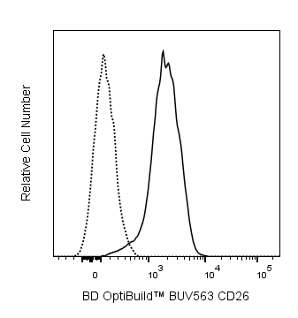
The H194-112 monoclonal antibody specifically binds to CD26, which is also known as, Thymocyte-activating molecule (THAM), or dipeptidyl peptidase IV (DPP IV, Dpp4). CD26 is a ~220- kDa dimer formed of identical type-II transmembrane core polypeptides which undergo variable post-translational modifications. It is a multi-functional molecule with both ectopeptidase and signal-transducing activities. Studies with specific DPP IV inhibitors suggest that the enzymatic activity is involved in the mediation of T-cell activation events. The expression of CD26 is developmentally regulated in the thymus. Resting lymphoid cells of the bone marrow and peripheral B and T lymphocytes express low levels of CD26; bone-marrow and peritoneal myeloid cells do not. CD26 is also found on epithelial cells in the kidney, liver, small intestine, and lung. Cross-linked H194-112 mAb induces proliferation of immature and mature thymocytes in the presence of either IL-1 plus IL-2 or PMA; addition of IL-2 or IL-4 to PMA further enhances the activation.
The antibody was conjugated to BD Horizon™ BUV563 which is part of the BD Horizon Brilliant™ Ultraviolet family of dyes. This dye is a tandem fluorochrome of BD Horizon BUV395 which has an Ex Max of 348 nm and an acceptor dye. The tandem has an Em Max at 563 nm. BD Horizon BUV563 can be excited by the 355 nm ultraviolet laser. On instruments with a 561 nm Yellow-Green laser, the recommended bandpass filter is 585/15 nm with a 535 nm long pass to minimize laser light leakage. When BD Horizon BUV563 is used with an instrument that does not have a 561 nm laser, a 560/40 nm filter with a 535 nm long pass may be more optimal. Due to the excitation and emission characteristics of the acceptor dye, there may be spillover into the PE and PE-CF594 detectors. However, the spillover can be corrected through compensation as with any other dye combination.

Development References (7)
-
Fleischer B. CD26: a surface protease involved in T-cell activation. Immunol Today. 1994; 15(4):180-184. (Biology). View Reference
-
Gorvel JP, Vivier I, Naquet P, Brekelmans P, Rigal A, Pierres M. Characterization of the neutral aminopeptidase activity associated to the mouse thymocyte-activating molecule. J Immunol. 1990; 144(8):2899-2907. (Biology). View Reference
-
Marguet D, Bernard AM, Vivier I, Darmoul D, Naquet P, Pierres M. cDNA cloning for mouse thymocyte-activating molecule. A multifunctional ecto-dipeptidyl peptidase IV (CD26) included in a subgroup of serine proteases. J Biol Chem. 1992; 267(4):2200-2208. (Biology). View Reference
-
Naquet P, MacDonald HR, Brekelmans P. A novel T cell-activating molecule (THAM) highly expressed on CD4-CD8- murine thymocytes. J Immunol. 1988; 141(12):4101-4109. (Immunogen: Activation). View Reference
-
Naquet P, Vivier I, Gorvel JP. Activation of mouse T lymphocytes by a monoclonal antibody to a developmentally regulated surface aminopeptidase (THAM). Immunol Rev. 1989; 111:177-193. (Clone-specific: Activation). View Reference
-
Reinhold D, Bank U, Buhling F. Inhibitors of dipeptidyl peptidase IV (DP IV, CD26) induces secretion of transforming growth factor-beta 1 (TGF-beta 1) in stimulated mouse splenocytes and thymocytes. Immunol Lett. 1997; 58(1):29-35. (Biology: Activation). View Reference
-
Vivier I, Marguet D, Naquet P . Evidence that thymocyte-activating molecule is mouse CD26 (dipeptidyl peptidase IV). J Immunol. 1988; 147(2):447-454. (Biology). View Reference
Please refer to Support Documents for Quality Certificates
Global - Refer to manufacturer's instructions for use and related User Manuals and Technical data sheets before using this products as described
Comparisons, where applicable, are made against older BD Technology, manual methods or are general performance claims. Comparisons are not made against non-BD technologies, unless otherwise noted.
For Research Use Only. Not for use in diagnostic or therapeutic procedures.