

Preparation And Storage
Product Notices
- Since applications vary, each investigator should titrate the reagent to obtain optimal results.
- An isotype control should be used at the same concentration as the antibody of interest.
- Caution: Sodium azide yields highly toxic hydrazoic acid under acidic conditions. Dilute azide compounds in running water before discarding to avoid accumulation of potentially explosive deposits in plumbing.
- The Alexa Fluor®, Pacific Blue™, and Cascade Blue® dye antibody conjugates in this product are sold under license from Molecular Probes, Inc. for research use only, excluding use in combination with microarrays, or as analyte specific reagents. The Alexa Fluor® dyes (except for Alexa Fluor® 430), Pacific Blue™ dye, and Cascade Blue® dye are covered by pending and issued patents.
- Alexa Fluor® is a registered trademark of Molecular Probes, Inc., Eugene, OR.
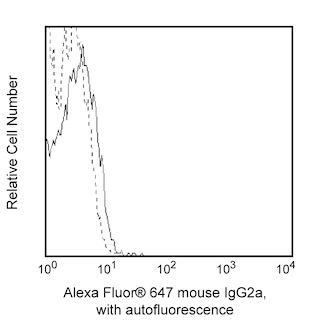
- Alexa Fluor® 647 fluorochrome emission is collected at the same instrument settings as for allophycocyanin (APC).
- For fluorochrome spectra and suitable instrument settings, please refer to our Multicolor Flow Cytometry web page at www.bdbiosciences.com/colors.
- Please refer to www.bdbiosciences.com/us/s/resources for technical protocols.
Companion Products


The human CD3 complex consists of four transmembrane proteins (γ, δ, ε, ζ) that are associated with the T cell antigen receptor (TCR) to form the CD3/TCR complex. The CD3 complex associates with either TCR αβ or TCR γδ heterodimers that are expressed by some thymocytes, T cells or NKT cells. The CD3 complex is required for the cell surface expression and signal-transducing functions of the TCR. The CD3 complex is expressed by ~60-85% thymocytes and ~60-80% of normal human peripheral blood lymphocytes. The MEM-57 monoclonal antibody reportedly recognizes the human CD3 epsilon subunit (CD3e/CD3ε, also known as T3E or TCRE) of the CD3/TCR complex. CD3e is a ~20 kDa unglycosylated transmembrane protein that belongs to the Ig gene superfamily. CD3e has an Ig-like extracellular domain (ECD) and an immunoreceptor tyrosine-based activation motif (ITAM) in its cytoplasmic domain. The MEM-57 antibody reportedly stimulates T cell proliferation and can induce cytotoxicity in some human T cell lines.
Development References (5)
-
Ernst DN, Shih CC. CD3 complex. J Biol Regul Homeost Agents. 2000; 14(3):226-229. (Biology). View Reference
-
Kurrle R. Cluster report: CD3. In: Knapp W. W. Knapp .. et al., ed. Leucocyte typing IV : white cell differentiation antigens. Oxford New York: Oxford University Press; 1989:290-293.
-
Pan Q, Brodeur JF, Drbal K, Dave VP. Different role for mouse and human CD3delta/epsilon heterodimer in preT cell receptor (preTCR) function: human CD3delta/epsilon heterodimer restores the defective preTCR function in CD3gamma- and CD3gammadelta-deficient mice.. Mol Immunol. 2006; 43(11):1741-50. (Clone-specific: Flow cytometry, Immunoprecipitation). View Reference
-
Transy C, Moingeon PE, Marshall B, Stebbins C, Reinherz EL. Most anti-human CD3 monoclonal antibodies are directed to the CD3 epsilon subunit.. Eur J Immunol. 1989; 19(5):947-50. (Clone-specific: Flow cytometry). View Reference
-
Tunnacliffe A, Olsson C, Traunecker A, Krissansen GW, Karjalainen K, de la Hera A. The majority of CD3 epitopes are conferred by the epsilon chain. In: Knapp W. W. Knapp .. et al., ed. Leucocyte typing IV : white cell differentiation antigens. Oxford New York: Oxford University Press; 1989:295-296.
Please refer to Support Documents for Quality Certificates
Global - Refer to manufacturer's instructions for use and related User Manuals and Technical data sheets before using this products as described
Comparisons, where applicable, are made against older BD Technology, manual methods or are general performance claims. Comparisons are not made against non-BD technologies, unless otherwise noted.
For Research Use Only. Not for use in diagnostic or therapeutic procedures.