
Preparation And Storage
Recommended Assay Procedures
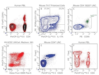
PE Mouse anti-FoxP1 antibody stained normal human peripheral blood B cells and T cells with a low signal-to-noise ratio (S/N ¡ 2) upon flow cytometric analyses. It is therefore not recommended for staining normal peripheral blood leucocytes for flow cytometric analysis.
Product Notices
- Since applications vary, each investigator should titrate the reagent to obtain optimal results.
- An isotype control should be used at the same concentration as the antibody of interest.
- Caution: Sodium azide yields highly toxic hydrazoic acid under acidic conditions. Dilute azide compounds in running water before discarding to avoid accumulation of potentially explosive deposits in plumbing.
- For fluorochrome spectra and suitable instrument settings, please refer to our Multicolor Flow Cytometry web page at www.bdbiosciences.com/colors.
- Species cross-reactivity detected in product development may not have been confirmed on every format and/or application.
- Cy is a trademark of GE Healthcare.
- Please refer to www.bdbiosciences.com/us/s/resources for technical protocols.
Companion Products


The JC12 monoclonal antibody specifically binds to Forkhead box protein P1 (FoxP1). FoxP1 belongs to subfamily P of the forkhead box (FOX) winged-helix transcription factor family. It plays a variety of roles such as regulating the formation of lymphoid, lung, brain and heart tissues. FoxP1 is critical in regulating early B cell development and is also required for maintenance of naïve T cell quiescence, and monocyte differentiation and macrophage function. Aberrant expression of FOXP1 has been linked with mucosa-associated lymphoid tissue lymphoma and diffuse-large B cell lymphoma.

Development References (10)
-
Banham AH, Beasley N, Campo E, et al. The FOXP1 winged helix transcription factor is a novel candidate tumor suppressor gene on chromosome 3p. Cancer Res. 2001; 61(24):8820-8829. (Immunogen: Immunohistochemistry, Western blot). View Reference
-
Brown PJ, Ashe SL, Leich E, et al. Potentially oncogenic B-cell activation-induced smaller isoforms of FOXP1 are highly expressed in the activated B cell-like subtype of DLBCL. Blood. 2008; 111(5):2816-2824. (Clone-specific: Immunohistochemistry, Western blot). View Reference
-
Feng X, Wang H, Takata H, Day TJ, Willen J, Hu H. Transcription factor Foxp1 exerts essential cell-intrinsic regulation of the quiescence of naive T cells. Nat Immunol. 2011; 12(6):544-550. (Biology). View Reference
-
Hu H, Wang B, Borde M, et al. Foxp1 is an essential transcriptional regulator of B cell development. Nat Immunol. 2006; 7(8):819-826. (Biology). View Reference
-
Katoh M, Igarashi M, Fukuda H, Nakagama H, Katoh M. Cancer genetics and genomics of human FOX family genes. Cancer Lett. 2013; 328(2):198-206. (Biology). View Reference
-
Sagardoy A, Martinez-Ferrandis JI, Roa S, et al. Downregulation of FOXP1 is required during germinal center B-cell function. Blood. 2013; 121(21):4311-4320. (Biology). View Reference
-
Shi C, Sakuma M, Mooroka T, et al. Down-regulation of the forkhead transcription factor Foxp1 is required for monocyte differentiation and macrophage function. Blood. 2008; 112(12):4699-4711. (Biology). View Reference
-
Streubel B, Vinatzer U, Lamprecht A, Raderer M, Chott A. T(3;14)(p14.1;q32) involving IGH and FOXP1 is a novel recurrent chromosomal aberration in MALT lymphoma. Leukemia. 2005; 19(4):652-658. (Biology). View Reference
-
Wang B, Lin D, Li C, Tucker P. Multiple domains define the expression and regulatory properties of Foxp1 forkhead transcriptional repressor. J Biol Chem. 2003; 278(27):24259-24268. (Biology). View Reference
-
Wlodarska I, Veyt E, De Paepe P, et al. FOXP1, a gene highly expressed in a subset of diffuse large B-cell lymphoma, is recurrently targeted by genomic aberrations. Leukemia. 2005; 19(8):1299-1305. (Clone-specific: Immunohistochemistry). View Reference
Please refer to Support Documents for Quality Certificates
Global - Refer to manufacturer's instructions for use and related User Manuals and Technical data sheets before using this products as described
Comparisons, where applicable, are made against older BD Technology, manual methods or are general performance claims. Comparisons are not made against non-BD technologies, unless otherwise noted.
For Research Use Only. Not for use in diagnostic or therapeutic procedures.