BD Pharmingen™ Purified Mouse anti-CRABP2
Clone M60-1228 (RUO)
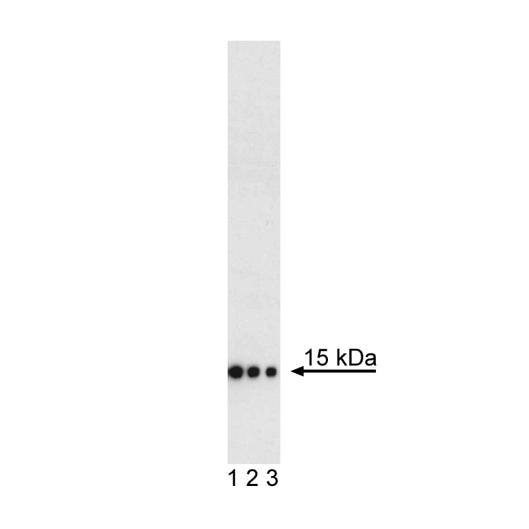
Western blot analysis of CRABP2 in human breast adenocarcinoma. MCF7 Cell Lysate (Cat. No. 611548) was probed with Purified Mouse anti-CRABP2 monoclonal antibody at titrations of 0.25 (lane 1), 0.05 (lane 2), and 0.01 µg/ml (lane 3). CRABP2 is identified as a band of 15 kDa.


Western blot analysis of CRABP2 in human breast adenocarcinoma. MCF7 Cell Lysate (Cat. No. 611548) was probed with Purified Mouse anti-CRABP2 monoclonal antibody at titrations of 0.25 (lane 1), 0.05 (lane 2), and 0.01 µg/ml (lane 3). CRABP2 is identified as a band of 15 kDa.
Preparation And Storage
Recommended Assay Procedures
Recommended Assay Procedure
1. Seed the cells in appropriate culture medium at an appropriate cell density in a BD Falcon™ 96-well Imaging Plate (Cat. No. 353219), and
culture overnight to 48 hours.
2. Remove the culture medium from the wells, and wash (one to two times) with 100 μl of 1× PBS.
3. Fix the cells by adding 100 µl of fresh 3.7% Formaldehyde in PBS or BD Cytofix™ fixation buffer (Cat. No. 554655) to each well and incubating for 10 minutes at room temperature (RT).
4. Remove the fixative from the wells, and wash the wells (one to two times) with 100 μl of 1× PBS.
5. Permeabilize the cells using either cold methanol (a), Triton™ X-100 (b), or Saponin (c):
a. Add 100 µl of -20°C 90% methanol or -20°C BD™ Phosflow Perm Buffer III (Cat. No. 558050) to each well and incubate for 5 minutes at RT.
b. Add 100 µl of 0.1% Triton™ X-100 to each well and incubate for 5 minutes at RT.
c. Add 100 µl of 1× Perm/Wash buffer (Cat. No. 554723) to each well and incubate for 15 to 30 minutes at RT. Continue to use 1× Perm/Wash buffer for all subsequent wash and dilutions steps.
6. Remove the permeabilization buffer from the wells, and wash one to two times with 100 μl of appropriate buffer (either 1× PBS or 1× Perm/Wash buffer, see step 5.c.).
7. Optional blocking step: Remove the wash buffers, and block the cells by adding 100 µl of blocking buffer BD Pharmingen™ Stain Buffer (FBS) (Cat. No. 554656) or 3% FBS in appropriate dilution buffer to each well and incubating for 15 to 30 minutes at RT.
8. Dilute the antibody to its optimal working concentration in appropriate dilution buffer. Titrate purified (unconjugated) antibodies and second-step reagents to determine the optimal concentration. If using a Bioimaging Certified antibody conjugate, dilute it 1:10.
9. Add 50 µl of diluted antibody per well and incubate for 60 minutes at RT. Incubate in the dark if using fluorescently labeled antibodies.
10. Remove the antibody, and wash the wells three times with 100 μl of wash buffer. An optional detergent wash (100 μl of 0.05% Tween in 1× PBS) can be included prior to the regular wash steps.
11. If the antibody being used is fluorescently labeled, then move to step 12. Otherwise, if using a purified unlabeled antibody, repeat steps 8 to 10 with a fluorescently labeled second-step reagent to detect the purified antibody.
12. After the final wash, counter-stain the nuclei by adding 100 μl of a 2 μg/ml solution of Hoechst 33342 (eg, Sigma-Aldrich Cat. No. B2261) in 1× PBS to each well at least 15 minutes before imaging.
13. View and analyze the cells on an appropriate imaging instrument.
Product Notices
- Since applications vary, each investigator should titrate the reagent to obtain optimal results.
- Please refer to www.bdbiosciences.com/us/s/resources for technical protocols.
- Caution: Sodium azide yields highly toxic hydrazoic acid under acidic conditions. Dilute azide compounds in running water before discarding to avoid accumulation of potentially explosive deposits in plumbing.
- Triton is a trademark of the Dow Chemical Company.
Companion Products


The M60-1228 monoclonal antibody reacts with Cellular Retinoic Acid-Binding Protein 2 (CRABP2). CRABP2 is a cytosolic low-molecular-weight protein with high affinity for retinoic acid (RA), a member of the vitamin A family that is able to induce cell differentiation. The translocation of CRABP2 to the nucleus indicates that it may function to deliver RA to RA receptors, which activate gene transcription. The differential expression of the CRABP2 gene during development and its overexpression in many cancers suggest that it is important in RA-mediated regulation of cell growth and differentiation.
Development References (2)
-
Gupta A, Williams BRG, Hanash SM. Rawwas J. Cellular retinoic acid-binding protein II is a direct transcriptional target of MycN in neuroblastoma. Cancer Res. 2006; 66(16):8100-8108. (Biology).
-
Lane MA, Xu J, Wilen EW, Sylvester R, Derguini F, Gudas LJ. LIF removal increases CRABP! and CRABPII transcripts in embryonic stem cells cultured in retinol or 4-oxoretinol. Mol Cell Endocrinol. 2008; 280(1-2):63-74. (Biology).
Please refer to Support Documents for Quality Certificates
Global - Refer to manufacturer's instructions for use and related User Manuals and Technical data sheets before using this products as described
Comparisons, where applicable, are made against older BD Technology, manual methods or are general performance claims. Comparisons are not made against non-BD technologies, unless otherwise noted.
For Research Use Only. Not for use in diagnostic or therapeutic procedures.