BD Pharmingen™ Purified Mouse Anti-Human SSEA-5
Clone 8e11/SSEA-5 (also known as 8e11) (RUO)
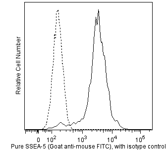
Flow cytometric analysis of SSEA-5 expression on human embryonic stem (ES) cells. H9 human ES cells (WiCell, Madison, WI) passage 48 grown in mTESR™1 media (StemCell Technologies) on BD Matrigel™ hESC-qualified Matrix (Cat. No. 354277) were harvested with Accutase™ Cell Detachment Solution (Cat. No. 561527) and stained with either Purified Mouse Anti-Human SSEA-5 antibody (solid line histogram) or with Purified Mouse IgG1, κ Isotype Control (Cat. No. 554121; dashed line histogram). The second-step reagent was FITC Goat Anti-Mouse Ig (Cat. No. 554001). The fluorescence histograms were derived from events with the forward and side light-scatter characteristics of the H9 human ES cell line. Flow cytometry was performed using a BD LSRFortessa™ Flow Cytometer System.
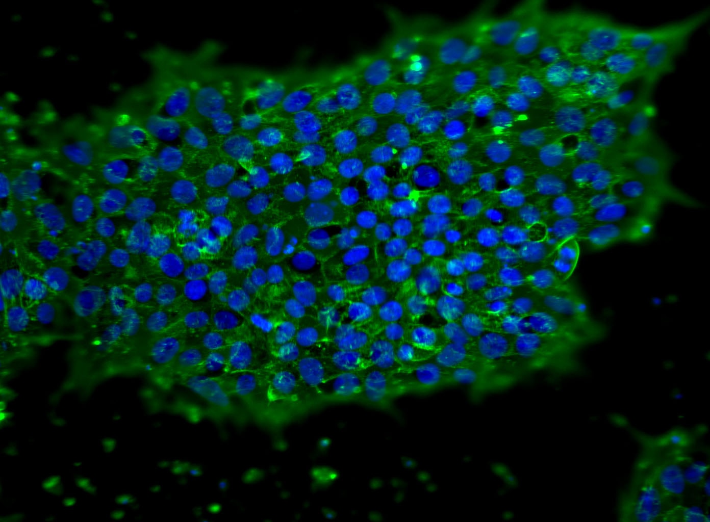
Immunoflourescent staining of SSEA-5 on human embryonic stem (ES) cells. H9 human ES cells (WiCell, Madison, WI) passage 43 grown in mTESR™1 media (StemCell Technologies) on BD Matrigel™ hESC-
qualified Matrix (Cat. No. 354277) were fixed with BD Cytofix™ Fixation Buffer (Cat. No. 554655). Cells were stained with Purified Mouse Anti-Human SSEA-5 monoclonal antibody (pseudo-colored green)at 5 μg/mL. The second-step reagent was Alexa Fluor® 488 Goat Anti-Mouse Ig (Life Technologies), and cell nuclei were stained with DAPI (pseudo-colored blue). The images were captured on a BD Pathway™ 435 Cell Analyzer and merged using BD Attovision™ software.

Flow cytometric analysis of SSEA-5 expression on human embryonic stem (ES) cells. H9 human ES cells (WiCell, Madison, WI) passage 48 grown in mTESR™1 media (StemCell Technologies) on BD Matrigel™ hESC-qualified Matrix (Cat. No. 354277) were harvested with Accutase™ Cell Detachment Solution (Cat. No. 561527) and stained with either Purified Mouse Anti-Human SSEA-5 antibody (solid line histogram) or with Purified Mouse IgG1, κ Isotype Control (Cat. No. 554121; dashed line histogram). The second-step reagent was FITC Goat Anti-Mouse Ig (Cat. No. 554001). The fluorescence histograms were derived from events with the forward and side light-scatter characteristics of the H9 human ES cell line. Flow cytometry was performed using a BD LSRFortessa™ Flow Cytometer System.
Immunoflourescent staining of SSEA-5 on human embryonic stem (ES) cells. H9 human ES cells (WiCell, Madison, WI) passage 43 grown in mTESR™1 media (StemCell Technologies) on BD Matrigel™ hESC-
qualified Matrix (Cat. No. 354277) were fixed with BD Cytofix™ Fixation Buffer (Cat. No. 554655). Cells were stained with Purified Mouse Anti-Human SSEA-5 monoclonal antibody (pseudo-colored green)at 5 μg/mL. The second-step reagent was Alexa Fluor® 488 Goat Anti-Mouse Ig (Life Technologies), and cell nuclei were stained with DAPI (pseudo-colored blue). The images were captured on a BD Pathway™ 435 Cell Analyzer and merged using BD Attovision™ software.
Preparation And Storage
Product Notices
- Since applications vary, each investigator should titrate the reagent to obtain optimal results.
- An isotype control should be used at the same concentration as the antibody of interest.
- Sodium azide is a reversible inhibitor of oxidative metabolism; therefore, antibody preparations containing this preservative agent must not be used in cell cultures nor injected into animals. Sodium azide may be removed by washing stained cells or plate-bound antibody or dialyzing soluble antibody in sodium azide-free buffer. Since endotoxin may also affect the results of functional studies, we recommend the NA/LE (No Azide/Low Endotoxin) antibody format, if available, for in vitro and in vivo use.
- Caution: Sodium azide yields highly toxic hydrazoic acid under acidic conditions. Dilute azide compounds in running water before discarding to avoid accumulation of potentially explosive deposits in plumbing.
- mTESR™1 is a trademark of StemCell Technologies.
- Accutase is a registered trademark of Innovative Cell Technologies, Inc.
- Patent Pending.
- Please refer to www.bdbiosciences.com/us/s/resources for technical protocols.
Companion Products


Stage-specific embryonic antigen (SSEA)-5 is a pluripotency surface marker expressed in the blastocyst inner cell mass and on human pluripotent stem cells, including both human embryonic stem cells (hESCs) and human induced pluripotent stem cells (hiPSCs). Because SSEA-5 expression rapidly decreases upon differentiation, it can be used to identify undifferentiated pluripotent stem cells. SSEA-5 can be combined with other pluripotency surface markers (e.g. CD9/CD90 or CD50/CD200) to immunodeplete remaining pluripotent stem cells from incompletely differentiated cultures.
Development References (1)
-
Tang C, Lee AS, Volkmer JP, et al. An antibody against SSEA-5 glycan on human pluripotent stem cells enables removal of teratoma-forming cells. Nat Biotechnol. 2011. (Clone-specific: Depletion, Flow cytometry, Immunofluorescence). View Reference
Please refer to Support Documents for Quality Certificates
Global - Refer to manufacturer's instructions for use and related User Manuals and Technical data sheets before using this products as described
Comparisons, where applicable, are made against older BD Technology, manual methods or are general performance claims. Comparisons are not made against non-BD technologies, unless otherwise noted.
For Research Use Only. Not for use in diagnostic or therapeutic procedures.