-
Reagents
- Flow Cytometry Reagents
-
Western Blotting and Molecular Reagents
- Immunoassay Reagents
-
Single-Cell Multiomics Reagents
- BD® OMICS-Guard Sample Preservation Buffer
- BD® AbSeq Assay
- BD® Single-Cell Multiplexing Kit
- BD Rhapsody™ ATAC-Seq Assays
- BD Rhapsody™ Whole Transcriptome Analysis (WTA) Amplification Kit
- BD Rhapsody™ TCR/BCR Next Multiomic Assays
- BD Rhapsody™ Targeted mRNA Kits
- BD Rhapsody™ Accessory Kits
- BD® OMICS-One Protein Panels
-
Functional Assays
-
Microscopy and Imaging Reagents
-
Cell Preparation and Separation Reagents
-
Training
- Flow Cytometry Basic Training
-
Product-Based Training
- FACSAria Product Based Training
- FACSMelody Product-Based Training
- FACSLyric Product-Based Training
- FACSCanto Product-Based Training
- LSRFortessa Product-Based Training
- FACSymphony Product-Based Training
- FACSDuet Product-Based Training
- HTS Product-Based Training
- BD FACSDiscover™ S8 Cell Sorter Product Training
-
Advanced Training
-
- BD® OMICS-Guard Sample Preservation Buffer
- BD® AbSeq Assay
- BD® Single-Cell Multiplexing Kit
- BD Rhapsody™ ATAC-Seq Assays
- BD Rhapsody™ Whole Transcriptome Analysis (WTA) Amplification Kit
- BD Rhapsody™ TCR/BCR Next Multiomic Assays
- BD Rhapsody™ Targeted mRNA Kits
- BD Rhapsody™ Accessory Kits
- BD® OMICS-One Protein Panels
-
- FACSAria Product Based Training
- FACSMelody Product-Based Training
- FACSLyric Product-Based Training
- FACSCanto Product-Based Training
- LSRFortessa Product-Based Training
- FACSymphony Product-Based Training
- FACSDuet Product-Based Training
- HTS Product-Based Training
- BD FACSDiscover™ S8 Cell Sorter Product Training
- Singapore (English)
-
Change country/language
Old Browser
This page has been recently translated and is available in French now.
Looks like you're visiting us from United States.
Would you like to stay on the current country site or be switched to your country?
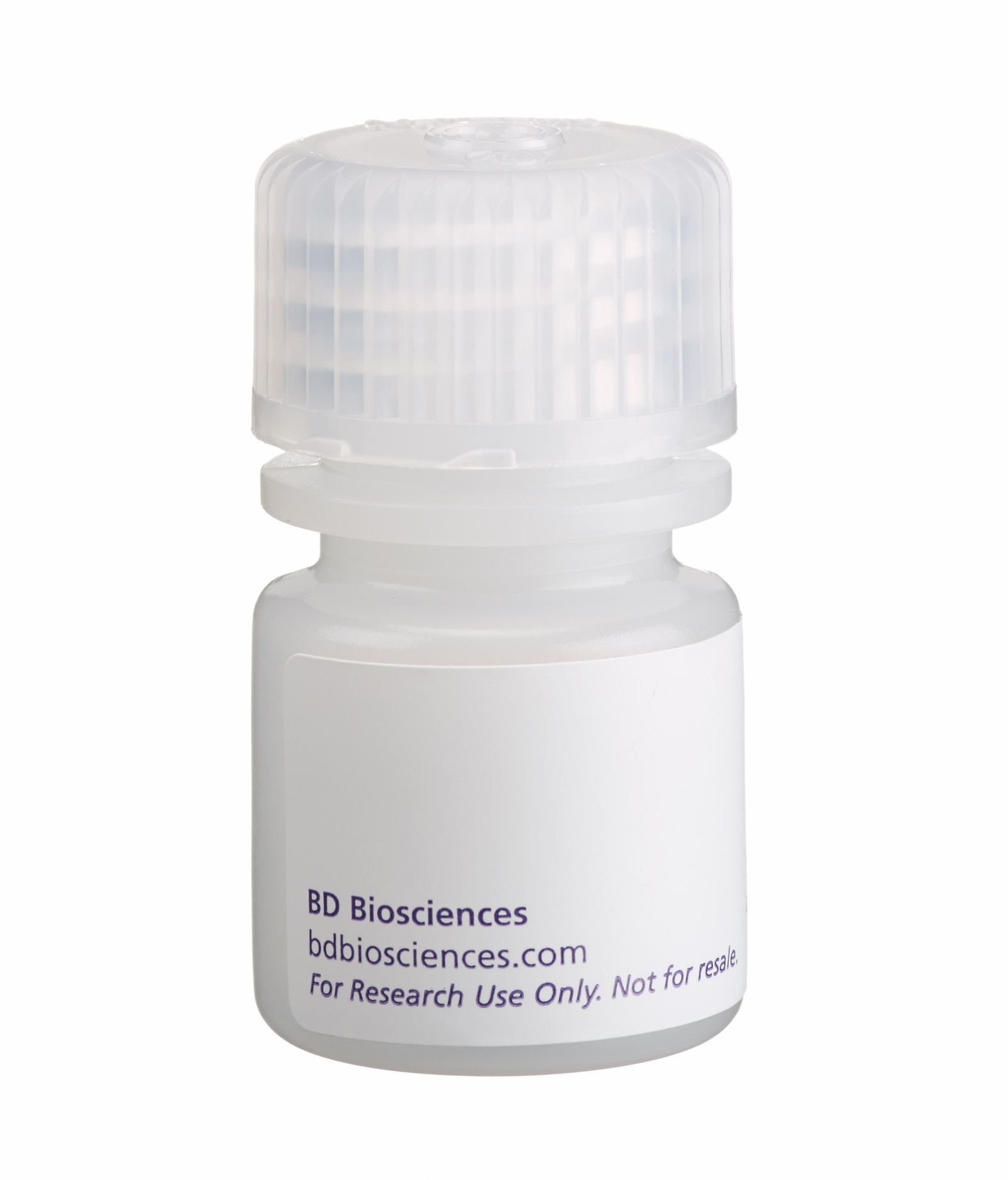
BD IMag™ Anti-Rat CD8 Magnetic Particles - DM
Clone G28 (RUO)

Positive selection of rat CD8+ T lymphocytes from rat spleen cells. Splenocytes were labeled with BD IMag™ anti-rat CD8 Particles - DM as described in the protocol. After labeling, the cells were separated using the BD IMagnet™, and the negative (CD8-) and positive (CD8+) fractions were collected. Please refer to the Separation Flow Chart to identify the separated cell populations represented in this figure. For flow cytometric analysis, fresh splenocytes (left panel), the negative fraction (middle panel), and the positive fraction (right panel) were stained with PE conjugated mouse anti-rat CD8 mAb OX-8 (Cat. no. 559976) and FITC-conjugated mouse anti-rat CD45 mAb OX-1 (Cat. no. 554877). The percent CD8+/CD45+ cells in each sample is given.


Positive selection of rat CD8+ T lymphocytes from rat spleen cells. Splenocytes were labeled with BD IMag™ anti-rat CD8 Particles - DM as described in the protocol. After labeling, the cells were separated using the BD IMagnet™, and the negative (CD8-) and positive (CD8+) fractions were collected. Please refer to the Separation Flow Chart to identify the separated cell populations represented in this figure. For flow cytometric analysis, fresh splenocytes (left panel), the negative fraction (middle panel), and the positive fraction (right panel) were stained with PE conjugated mouse anti-rat CD8 mAb OX-8 (Cat. no. 559976) and FITC-conjugated mouse anti-rat CD45 mAb OX-1 (Cat. no. 554877). The percent CD8+/CD45+ cells in each sample is given.

Positive selection of rat CD8+ T lymphocytes from rat spleen cells. Splenocytes were labeled with BD IMag™ anti-rat CD8 Particles - DM as described in the protocol. After labeling, the cells were separated using the BD IMagnet™, and the negative (CD8-) and positive (CD8+) fractions were collected. Please refer to the Separation Flow Chart to identify the separated cell populations represented in this figure. For flow cytometric analysis, fresh splenocytes (left panel), the negative fraction (middle panel), and the positive fraction (right panel) were stained with PE conjugated mouse anti-rat CD8 mAb OX-8 (Cat. no. 559976) and FITC-conjugated mouse anti-rat CD45 mAb OX-1 (Cat. no. 554877). The percent CD8+/CD45+ cells in each sample is given.


BD IMag™ Anti-Rat CD8 Magnetic Particles - DM

BD IMag™ Anti-Rat CD8 Magnetic Particles - DM

Regulatory Status Legend
Any use of products other than the permitted use without the express written authorization of Becton, Dickinson and Company is strictly prohibited.
Preparation And Storage
Recommended Assay Procedures
MAGNETIC LABELING PROTOCOL
1. Prepare a single-cell suspension from the lymphoid tissue of interest according to standard laboratory procedures. Remove clumps of cells and/or debris by passing the suspension through a 70-mm nylon cell strainer.
2. Dilute BD IMag™ Buffer (10X) (Cat. no. 552362) 1:10 with sterile distilled water or prepare 1X BD IMag™ buffer by supplementing Phosphate Buffered Saline with 0.5% BSA, 2 mM EDTA, and 0.09% sodium azide. Store at 4˚C.
3. Wash cells with an excess volume of 1X BD IMag™ buffer, and carefully aspirate all the supernatant.
4. Vortex the BD IMag™ anti-rat CD8 Particles - DM thoroughly, and add 50 μl of particles for every 10 million total cells.
5. MIX THOROUGHLY. Refrigerate at 6°C to 12°C for 30 minutes.
6. Bring the BD IMag-particle labeling volume up to 10 to 80 million cells/ml with 1X BD IMag™ buffer, and immediately place the tube on the BD IMagnet™. Incubate at room temperature for 8-10 minutes.
7. With the tube on the BD IMagnet™, carefully pipette off the supernatant. This supernatant contains the negative fraction.
8. Remove the tube from the BD IMagnet™, and add 1 ml of 1X BD IMag™ buffer. Gently resuspend cells by pipetting up and down, and return the tube to the BD IMagnet™ for another 2 - 4 minutes.
9. With the tube on the BD IMagnet™, carefully pipette off the supernatant and discard.
10. Repeat Steps 8 and 9.
11. After the final wash step, resuspend the positive fraction in an appropriate buffer or media, and proceed with desired downstream application(s).
NOTE: The concentration of BD IMag™ anti-rat CD8 Particles - DM suggested in the protocol has been optimized for the purification of CD8-positive T lymphocytes from rat splenocytes. When labeling target cell populations present at lower frequencies, fewer BD IMag™ particles can be used. Conversely, when labeling target cell populations that are present at higher frequencies, more particles should be used. To determine the optimal concentration of the BD IMag™ anti-rat CD8 Particles - DM for a particular application, a titration in two-fold increments is recommended.
NOTE: Avoid nonspecific labeling by working quickly and keeping incubation times as recommended.
Product Notices
- Since applications vary, each investigator should titrate the reagent to obtain optimal results.
- BD IMag™ particles are prepared from carboxy-functionalized magnetic particles which are manufactured by Skold Technology and are licensed under US patent number 7,169,618.
- Caution: Sodium azide yields highly toxic hydrazoic acid under acidic conditions. Dilute azide compounds in running water before discarding to avoid accumulation of potentially explosive deposits in plumbing.
- Source of all serum proteins is from USDA inspected abattoirs located in the United States.
- Please refer to www.bdbiosciences.com/us/s/resources for technical protocols.
BD IMag™ anti-rat CD8 Magnetic Particles - DM are magnetic nanoparticles that have monoclonal antibody conjugated to their surfaces. These particles are optimized for the positive selection or depletion of CD8-bearing T lymphocytes using the BD IMagnet™. The G28 antibody reacts with the V-like domain of the α chain of the CD8 differentiation antigen. CD8α is an approximately 32-34 kDa cell surface receptor expressed either as a heterodimer with the CD8 ß chain (CD8 αß) or as a homodimer (CD8 αα). The CD8 αß heterodimer (CD8a and CD8b, respectively) is located on the surface of most thymocytes and a subpopulation of mature T lymphocytes (ie, MHC class I-restricted T cells, including most T suppressor/cytotoxic cells). Intestinal intraepithelial lymphocytes, many CD8+ T cells of athymic rats, many activated CD4+ T cells, and most NK cells express CD8a without CD8b. It has been suggested that the expression of the CD8a/CD8b heterodimer is restricted to thymus-derived T lymphocytes. CD8a reportedly is not expressed on resting CD4+ T-helper cells.
A single-cell suspension from the lymphoid tissue of interest is labeled with BD IMag™ anti-rat CD8 Magnetic Particles - DM according to the following Magnetic Labeling Protocol. This labeled cell suspension is then placed within the magnetic field of the BD IMagnet™ (Cat. no. 552311). Labeled cells migrate toward the magnet (positive fraction), leaving the unlabeled cells in suspension so they can be drawn off (negative fraction). The tube is then removed from the magnetic field for resuspension of the positive fraction. The separation is repeated twice to increase the purity of the positive fraction. The magnetic separation steps are diagrammed in the Separation Flow Chart. After the positive fraction is washed, the small size of the magnetic particles allows the positive fraction to be further evaluated in downstream applications such as flow cytometry.
Development References (3)
-
Janeway CA Jr. The T cell receptor as a multicomponent signalling machine: CD4/CD8 coreceptors and CD45 in T cell activation. Annu Rev Immunol. 1992; 10:645-674. (Biology). View Reference
-
Mitnacht R, Bischof A, Torres-Nagel N, Hunig T. Opposite CD4/CD8 lineage decisions of CD4+8+ mouse and rat thymocytes to equivalent triggering signals: correlation with thymic expression of a truncated CD8 alpha chain in mice but not rats. J Immunol. 1998; 160(2):700-707. (Biology). View Reference
-
Torres-Nagel N, Kraus E, Brown MH, et al. Differential thymus dependence of rat CD8 isoform expression.. Eur J Immunol. 1992; 22(11):2841-2848. (Biology). View Reference
Please refer to Support Documents for Quality Certificates
Global - Refer to manufacturer's instructions for use and related User Manuals and Technical data sheets before using this products as described
Comparisons, where applicable, are made against older BD Technology, manual methods or are general performance claims. Comparisons are not made against non-BD technologies, unless otherwise noted.
For Research Use Only. Not for use in diagnostic or therapeutic procedures.