A Guide to Flow Cytometry Markers
August 30, 2023
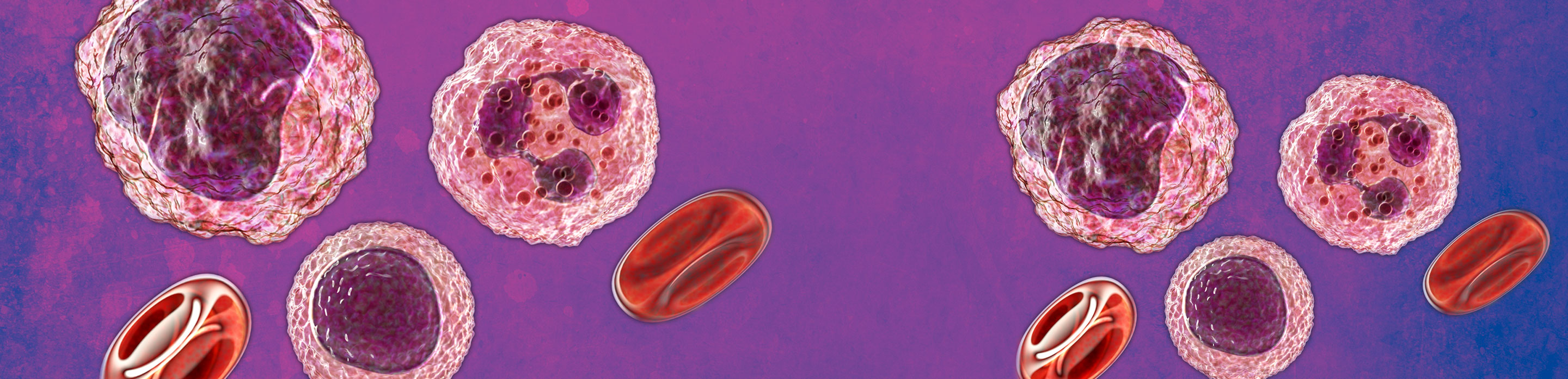
Classically, host immunity is divided into innate and adaptive immune responses.1 Innate immune cells include myeloid cells (monocytes, macrophages, granulocytes and dendritic cells), natural killer (NK) cells and innate lymphoid cells. Adaptive immune cells include the immunoglobulin family and cells such as B- and T-lymphocytes.1,2
Immunophenotyping utilises the unique ability of flow cytometry to simultaneously analyse mixed populations of immune cells through the presence or absence of multiple flow cytometry markers.3,4
It allows for the characterisation of cell populations and subpopulations, identification of the status of cell differentiation and quantification of surface or intracellular molecules associated with specific cellular functions.3
Flow cytometry markers lists
Most antigens are given "cluster of differentiation" (CD) numbers by the Human Leukocyte Differentiation Workshops, so that a common nomenclature is used to define monoclonal antibodies that are directed against specific cellular antigens.4 For example, CD3 is "cluster of differentiation number 3" and is used to define the T-cell co-receptor that is present on all T cells.4
Most immune cells have specific CD markers that define them as a population of cells.1 These cell markers are called lineage markers and are used to define specific cell populations for additional analysis in each immunophenotyping experiment.4
As well as lineage markers that define populations of immune cells, other markers are used to characterise each cell population, such as activation markers (CD69 CD25, and CD62L).4
Myeloid cell markers
For phenotypic identification of myeloid cell populations, markers CD33, CD123, CD14, CD16, HLA-DR, CD11c, CD141 and CD1c can be used.5 One study developed a 10-colour flow cytometry protocol for quantification of myeloid cells, monocytes and granulocytes.6
| 10-color flow cytometric protocols for quantification of all major leukocyte populations6 | |
| Myeloid cells | LIN2, CD123, HLA-DR, CD11c, CD11b, CD33, CD16 |
| Monocytes-1 | CD80, CD143, CD14, CD32, CD64, CD86, CD16, HLA-DR, CD45 |
| Monocytes-2 | B7H1, TNFR2, CD14, PD-1, CD40, CD16, HLA-DR, CD45 |
| Granulocytes | CD66b, CD63, CD14, CD44, CD203c, CCR3, CD16, CD49d, CD15, CD45 |
CD14, CD163 and CD68 can detect macrophages in human lung tumours, with CD163 being a macrophage-specific marker.7 In human synovial fluid, CD11c and CD206 identified M1 and M2 macrophages, respectively.8 CD38 is also a highly and consistently induced inflammatory marker in human macrophage responses, so may play a role in monitoring inflammatory disease status.9
There are three major dendritic cell (DC) subsets: plasmacytoid DC (pDC), myeloid/conventional DC1 (cDC1) and myeloid/conventional DC2 (cDC2).10 Original DC markers of CD141 and CD1c have limitations as both are induced on cDC and monocyte-derived cells in tissues and in culture.10
| Myeloid cell marker10 | Function |
|---|---|
| CD15 | Granulocyte subsets |
| CD66b | |
| CD16 | |
| CD45 | |
| FCeR1a | |
| CD123 | |
| Siglec 8 | |
| CD14 | Monocyte subsets |
| HLA-DR | |
| CD45 | |
| CD16 | |
| CD32 | |
| CD64 | |
| HLA-DR | DC subsets |
| CD123 | |
| CD11c | |
| CD1c | |
| CD141 | |
| CD80 | Activation markers |
| CD86 |
Natural-killer cells
Common natural-killer (NK) cell markers are CD56 and CD161.4 CD56bright and CD56dim NK cells in peripheral blood were found to display an activated effector phenotype that is similar in nature to moderate and severe COVID-19 disease.11 NK-cell activation was assessed by analysing expression of Ki-67, HLA-DR (human leukocyte antigen-DR) and CD69.11
T-cell markers
CD4+ T-αβ helper cell subsets include Th1, Th2, Th9, Th17, Th22 and regulatory T cells (Tregs), while CD8+ T-αβ cell subsets include cytotoxic T lymphocytes (Tc)1, Tc2, Tc9, Tc17 and CD8+ Tregs.12
| T-Cell marker12 | Function |
|---|---|
| CD3 | Lineage markers |
| CD4 | |
| CD8 | |
| CD45 | |
| CCR4 | T-helper subsets |
| CCR6 | |
| CCR10 | |
| CXCR3 | |
| CXCR5 | |
| CD25 | Treg |
| CD127 | |
| FoxP3 | |
| CD45RA | Maturation |
| CD45RO | |
| CCR7 | |
| CD27 | |
| CD95 | |
| CD28 | |
| PD-1 | Exhaustion |
| CTLA-4 | |
| TGIT | |
| CD69 | Activation |
| HLA-DR | |
| CD107 | Cytotoxicity (upon activation) |
| Granzyme B | |
| Perforin |
Specifically, for Treg cells, CD3, CD4, CD25, CD127 and Foxp3 markers as the minimally required markers to define human Tregs to allow for robust and undisputable gating.13 Ki67 and CD45RA are highly recommended for recently activated/proliferated Tregs and discriminating between naïve and TCR-triggered Tregs, respectively.13
B-cell markers
B cells are identified as CD19pos and can be further classified as transitional, mature-naïve, memory, atypical memory, activated B cells and plasmablasts; flow cytometry markers allow recognition of these subsets.14
| B-cell marker14 | Function |
|---|---|
| CD19 | Lineage marker |
| CD20 | |
| CD27 | |
| IgD | |
| CD38 | T-helper subsets |
| IgM | |
| IgG | |
| CD24 |