Skip to main content Skip to navigation
Biotin Mouse Anti-Human MCP-1
ELISA standard curve of recombinant human MCP-1. Sandwich ELISA was performed according to ELISA detection protocol described below in the recommended assay procedure.
Product Details
Down Arrow Up Arrow


BD Pharmingen™
CCL2; C-C motif chemokine 2; Chemokine (C-C motif) ligand 2; MCAF; SCYA2
Human (QC Testing), Rhesus, Cynomolgus (Tested in Development)
Mouse IgG1, κ
Recombinant Human MCP-1
ELISA Detection (Routinely Tested)
0.5 mg/ml
AB_395498
Aqueous buffered solution containing ≤0.09% sodium azide.
RUO


Preparation And Storage

Store undiluted at 4°C. The monoclonal antibody was purified from tissue culture supernatant or ascites by affinity chromatography. The antibody was conjugated with biotin under optimum conditions, and unreacted biotin was removed.

Recommended Assay Procedures

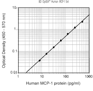
ELISA Detection: Biotin Mouse Anti-Human MCP-1 (Cat. No. 554664) is useful as a detection antibody for a sandwich ELISA for measuring human MCP-1 protein levels.  Biotin Mouse Anti-Human MCP-1 can be paired with Purified Mouse Anti-Human MCP-1 (clone 10F7.2; Cat. No. 555055) as the capture antibody and recombinant human MCP-1 (Cat. No. 554620) as the standard. The Biotin Mouse Anti-Human MCP-1 should be titrated 0.5 - 4 µg/ml to determine the optimal concentration for ELISA detection. To obtain linear standard curves, doubling dilutions of human MCP-1 ranging from ~5,000 to 15 pg/ml are recommended for inclusion in each ELISA plate. For specific methodology, please visit the protocols section under "ELISA and ELISPOT" on our web site at http://www.bdbiosciences.com/us/s/resources.

Note 1: This ELISA pair shows no cross-reactivity with any of the following chemokines tested ( e.g., Human eotaxin , MCP-2, MCP-3, MCP-4, mouse MCP-1 and rat MCP-1).

Note 2: This ELISA pair is recommended primarily for measuring cytokine from experimental cell culture systems. These ELISA reagents are not recommended for assaying serum or plasma samples. For measurement of human MCP-1 in serum or plasma samples the BD OptEIA™ Human MCP-1 ELISA Set (Cat. No. 555179) or the BD OptEIA™ Human MCP-1 ELISA Kit (Cat. No. 559017) are recommended.

Product Notices

  1. Since applications vary, each investigator should titrate the reagent to obtain optimal results.
  2. Caution: Sodium azide yields highly toxic hydrazoic acid under acidic conditions. Dilute azide compounds in running water before discarding to avoid accumulation of potentially explosive deposits in plumbing.
  3. For fluorochrome spectra and suitable instrument settings, please refer to our Multicolor Flow Cytometry web page at www.bdbiosciences.com/colors.
  4. Species cross-reactivity detected in product development may not have been confirmed on every format and/or application.
  5. Please refer to www.bdbiosciences.com/us/s/resources for technical protocols.
554664 Rev. 3
Antibody Details
Down Arrow Up Arrow
5D3-F7

The 5D3-F7 monoclonal antibody specifically binds to human monocyte chemoattractant protein-1 (MCP-1), also known as CCL2 (C-C motif chemokine 2), Monocyte chemotactic and activating factor (MCAF), and Small-inducible cytokine A2 (SCYA2). MCP-1 is a 10-14 kDa glycoprotein member of the beta or CC family of chemokines. It expressed by monocytes, fibroblasts, endothelial cells and other cell types in response to IL-1, IL-6, TNF, and a variety of other stimuli. MCP-1 binds to and exerts its biological activity through G-protein coupled chemokine receptors including CCR2/CD192 and CCR4/CD194. It serves as a chemoattractant and activating factor for monocytes and other cell types including T cells, NK cells, and basophils.

MCP-1 is a member of the CC chemokine family and it is produced by monocytes, T lymphocytes, fibroblasts, endothelial cells, smooth muscle cells, keratinocytes and some tumors. Its production can be induced by LPS or IFN-γ. Clone 5D3-F7 also cross reacts with an intracellular component of LPS-stimulated (24 hours) peripheral blood monocytes of rhesus and cynomolgus macaque monkeys. The staining pattern observed on non-human primate monocytes is not as strong as that seen on normal human peripheral blood monocytes.

554664 Rev. 3
Format Details
Down Arrow Up Arrow
Biotin
Biotin is a ubiquitous co-factor (also known as Vitamin B7) that has many properties that make it extremely useful for molecular biology. Biotin has an extremely high affinity for the Avidin family of proteins (Kd = 10-15 M), making it the perfect tool to link two molecules. Biotin labeled antibodies can be combined with any number of Avidin-conjugated probes in order to customize an assay to a particular need. This is especially useful in the case of magnetic cell separation using streptavidin/magnetic bead conjugates, or in the case of flow cytometry using streptavidin/fluorophore conjugates.
Biotin
554664 Rev.3
Citations & References
Down Arrow Up Arrow

Development References (3)

  1. Matsushima K, Oppenheim JJ. Interleukin 8 and MCAF: novel inflammatory cytokines inducible by IL 1 and TNF. Cytokine. 1989; 1(1):2-13. (Clone-specific). View Reference
  2. Peri G, Milanese C, Matteucci C, et al. A new monoclonal antibody (5D3-F7) which recognizes human monocyte-chemotactic protein-1 but not related chemokines. Development of a sandwich ELISA and in situ detection of producing cells. J Immunol Methods. 1994; 174(1-2):249-257. (Clone-specific: ELISA, Neutralization, Western blot). View Reference
  3. Yoshimura T, Yuhki N, Moore SK, Appella E, Lerman MI, Leonard EJ. Human monocyte chemoattractant protein-1 (MCP-1). Full-length cDNA cloning, expression in mitogen-stimulated blood mononuclear leukocytes, and sequence similarity to mouse competence gene JE. FEBS Lett. 1989; 244(2):487-493. (Clone-specific). View Reference
554664 Rev. 3

Please refer to Support Documents for Quality Certificates


Global - Refer to manufacturer's instructions for use and related User Manuals and Technical data sheets before using this products as described


Comparisons, where applicable, are made against older BD Technology, manual methods or are general performance claims.  Comparisons are not made against non-BD technologies, unless otherwise noted.

For Research Use Only. Not for use in diagnostic or therapeutic procedures.