Dear Customer, If you experience any text readability or display issues on our website, please update your web browser to the latest version
.
See More
Dear Customer, If you experience any text readability or display issues on our website, please update your web browser to the latest version
.
See MoreDear Customer, If you experience any text readability or display issues on our website, please update your web browser to the latest version
.
This page has been recently translated and is available in French now.
Looks like you're visiting us from {countryName}.
Would you like to stay on the current location site or be switched to your location?
Two-color flow cytometric analysis of GM-CSF expression in stimulated mouse T cells. Mouse splenic CD4+ T cells were cultured (2 days) with plate-bound Purified NA/LE Hamster Anti-Mouse CD3e (Cat. No. 553057; 10 μg/ml for coating) and soluble Purified NA/LE Hamster Anti-Mouse CD28 (Cat. No. 553294; 2 μg/ml) antibodies with Recombinant Mouse IL-2 (Cat. No. 550069; 10 ng/ml) and Mouse IL-4 (Cat. No. 550067; 25 ng/ml). The cells were subsequently cultured (3 days) with Recombinant IL-2 and IL-4. The cells were then stimulated with PMA (Sigma Cat. No. P-8139) and Ionomycin (Sigma Cat. No. I-0634) in the presence of BD GolgiStop™ Protein Transport Inhibitor (containing Monensin) (Cat. No. 554724) for 6 hours.
The cells were harvested, washed, and fixed with BD Cytofix™ Fixation Buffer (Cat. No. 554655). The cells were then washed and stained in BD Perm/Wash™ Buffer (Cat. No. 554723) with APC Rat Anti-Mouse CD4 antibody (Cat. No. 553051/561091) and either BD Horizon™ BV421 Rat IgG2a, κ Isotype Control (Cat. No. 562602; Left Panel) or BD Horizon BV421 Rat Anti-Mouse GM-CSF antibody (Cat. No. 564747; Right Panel). Two-color flow cytometric dot plots showing correlated expression of GM-CSF (or Ig Isotype control staining) versus CD4 were derived from gated events with the forward and side light-scatter characteristics of intact stimulated lymphocytes. Flow cytometric analysis was performed using a BD™ LSR II Flow Cytometer System.
Two-color flow cytometric analysis of GM-CSF expression in stimulated mouse T cells. Mouse splenic CD4+ T cells were cultured (2 days) with plate-bound Purified NA/LE Hamster Anti-Mouse CD3e (Cat. No. 553057; 10 μg/ml for coating) and soluble Purified NA/LE Hamster Anti-Mouse CD28 (Cat. No. 553294; 2 μg/ml) antibodies with Recombinant Mouse IL-2 (Cat. No. 550069; 10 ng/ml) and Mouse IL-4 (Cat. No. 550067; 25 ng/ml). The cells were subsequently cultured (3 days) with Recombinant IL-2 and IL-4. The cells were then stimulated with PMA (Sigma Cat. No. P-8139) and Ionomycin (Sigma Cat. No. I-0634) in the presence of BD GolgiStop™ Protein Transport Inhibitor (containing Monensin) (Cat. No. 554724) for 6 hours.
The cells were harvested, washed, and fixed with BD Cytofix™ Fixation Buffer (Cat. No. 554655). The cells were then washed and stained in BD Perm/Wash™ Buffer (Cat. No. 554723) with APC Rat Anti-Mouse CD4 antibody (Cat. No. 553051/561091) and either BD Horizon™ BV421 Rat IgG2a, κ Isotype Control (Cat. No. 562602; Left Panel) or BD Horizon BV421 Rat Anti-Mouse GM-CSF antibody (Cat. No. 564747; Right Panel). Two-color flow cytometric dot plots showing correlated expression of GM-CSF (or Ig Isotype control staining) versus CD4 were derived from gated events with the forward and side light-scatter characteristics of intact stimulated lymphocytes. Flow cytometric analysis was performed using a BD™ LSR II Flow Cytometer System.
Two-color flow cytometric analysis of GM-CSF expression in stimulated mouse T cells. Mouse splenic CD4+ T cells were cultured (2 days) with plate-bound Purified NA/LE Hamster Anti-Mouse CD3e (Cat. No. 553057; 10 μg/ml for coating) and soluble Purified NA/LE Hamster Anti-Mouse CD28 (Cat. No. 553294; 2 μg/ml) antibodies with Recombinant Mouse IL-2 (Cat. No. 550069; 10 ng/ml) and Mouse IL-4 (Cat. No. 550067; 25 ng/ml). The cells were subsequently cultured (3 days) with Recombinant IL-2 and IL-4. The cells were then stimulated with PMA (Sigma Cat. No. P-8139) and Ionomycin (Sigma Cat. No. I-0634) in the presence of BD GolgiStop™ Protein Transport Inhibitor (containing Monensin) (Cat. No. 554724) for 6 hours.
The cells were harvested, washed, and fixed with BD Cytofix™ Fixation Buffer (Cat. No. 554655). The cells were then washed and stained in BD Perm/Wash™ Buffer (Cat. No. 554723) with APC Rat Anti-Mouse CD4 antibody (Cat. No. 553051/561091) and either BD Horizon™ BV421 Rat IgG2a, κ Isotype Control (Cat. No. 562602; Left Panel) or BD Horizon BV421 Rat Anti-Mouse GM-CSF antibody (Cat. No. 564747; Right Panel). Two-color flow cytometric dot plots showing correlated expression of GM-CSF (or Ig Isotype control staining) versus CD4 were derived from gated events with the forward and side light-scatter characteristics of intact stimulated lymphocytes. Flow cytometric analysis was performed using a BD™ LSR II Flow Cytometer System.
Two-color flow cytometric analysis of GM-CSF expression in stimulated mouse T cells. Mouse splenic CD4+ T cells were cultured (2 days) with plate-bound Purified NA/LE Hamster Anti-Mouse CD3e (Cat. No. 553057; 10 μg/ml for coating) and soluble Purified NA/LE Hamster Anti-Mouse CD28 (Cat. No. 553294; 2 μg/ml) antibodies with Recombinant Mouse IL-2 (Cat. No. 550069; 10 ng/ml) and Mouse IL-4 (Cat. No. 550067; 25 ng/ml). The cells were subsequently cultured (3 days) with Recombinant IL-2 and IL-4. The cells were then stimulated with PMA (Sigma Cat. No. P-8139) and Ionomycin (Sigma Cat. No. I-0634) in the presence of BD GolgiStop™ Protein Transport Inhibitor (containing Monensin) (Cat. No. 554724) for 6 hours.
The cells were harvested, washed, and fixed with BD Cytofix™ Fixation Buffer (Cat. No. 554655). The cells were then washed and stained in BD Perm/Wash™ Buffer (Cat. No. 554723) with APC Rat Anti-Mouse CD4 antibody (Cat. No. 553051/561091) and either BD Horizon™ BV421 Rat IgG2a, κ Isotype Control (Cat. No. 562602; Left Panel) or BD Horizon BV421 Rat Anti-Mouse GM-CSF antibody (Cat. No. 564747; Right Panel). Two-color flow cytometric dot plots showing correlated expression of GM-CSF (or Ig Isotype control staining) versus CD4 were derived from gated events with the forward and side light-scatter characteristics of intact stimulated lymphocytes. Flow cytometric analysis was performed using a BD™ LSR II Flow Cytometer System.
Two-color flow cytometric analysis of GM-CSF expression in stimulated mouse T cells. Mouse splenic CD4+ T cells were cultured (2 days) with plate-bound Purified NA/LE Hamster Anti-Mouse CD3e (Cat. No. 553057; 10 μg/ml for coating) and soluble Purified NA/LE Hamster Anti-Mouse CD28 (Cat. No. 553294; 2 μg/ml) antibodies with Recombinant Mouse IL-2 (Cat. No. 550069; 10 ng/ml) and Mouse IL-4 (Cat. No. 550067; 25 ng/ml). The cells were subsequently cultured (3 days) with Recombinant IL-2 and IL-4. The cells were then stimulated with PMA (Sigma Cat. No. P-8139) and Ionomycin (Sigma Cat. No. I-0634) in the presence of BD GolgiStop™ Protein Transport Inhibitor (containing Monensin) (Cat. No. 554724) for 6 hours.
The cells were harvested, washed, and fixed with BD Cytofix™ Fixation Buffer (Cat. No. 554655). The cells were then washed and stained in BD Perm/Wash™ Buffer (Cat. No. 554723) with APC Rat Anti-Mouse CD4 antibody (Cat. No. 553051/561091) and either BD Horizon™ BV421 Rat IgG2a, κ Isotype Control (Cat. No. 562602; Left Panel) or BD Horizon BV421 Rat Anti-Mouse GM-CSF antibody (Cat. No. 564747; Right Panel). Two-color flow cytometric dot plots showing correlated expression of GM-CSF (or Ig Isotype control staining) versus CD4 were derived from gated events with the forward and side light-scatter characteristics of intact stimulated lymphocytes. Flow cytometric analysis was performed using a BD™ LSR II Flow Cytometer System.
Two-color flow cytometric analysis of GM-CSF expression in stimulated mouse T cells. Mouse splenic CD4+ T cells were cultured (2 days) with plate-bound Purified NA/LE Hamster Anti-Mouse CD3e (Cat. No. 553057; 10 μg/ml for coating) and soluble Purified NA/LE Hamster Anti-Mouse CD28 (Cat. No. 553294; 2 μg/ml) antibodies with Recombinant Mouse IL-2 (Cat. No. 550069; 10 ng/ml) and Mouse IL-4 (Cat. No. 550067; 25 ng/ml). The cells were subsequently cultured (3 days) with Recombinant IL-2 and IL-4. The cells were then stimulated with PMA (Sigma Cat. No. P-8139) and Ionomycin (Sigma Cat. No. I-0634) in the presence of BD GolgiStop™ Protein Transport Inhibitor (containing Monensin) (Cat. No. 554724) for 6 hours.
The cells were harvested, washed, and fixed with BD Cytofix™ Fixation Buffer (Cat. No. 554655). The cells were then washed and stained in BD Perm/Wash™ Buffer (Cat. No. 554723) with APC Rat Anti-Mouse CD4 antibody (Cat. No. 553051/561091) and either BD Horizon™ BV421 Rat IgG2a, κ Isotype Control (Cat. No. 562602; Left Panel) or BD Horizon BV421 Rat Anti-Mouse GM-CSF antibody (Cat. No. 564747; Right Panel). Two-color flow cytometric dot plots showing correlated expression of GM-CSF (or Ig Isotype control staining) versus CD4 were derived from gated events with the forward and side light-scatter characteristics of intact stimulated lymphocytes. Flow cytometric analysis was performed using a BD™ LSR II Flow Cytometer System.
Any use of products other than the permitted use without the express written authorization of Becton, Dickinson and Company is strictly prohibited.
The MP1-22E9 monoclonal antibody specifically binds to mouse Granulocyte-Macrophage Colony Stimulating Factor (GM-CSF). The immunogen used to generate the MP1-22E9 hybridoma was yeast-expressed recombinant mouse GM-CSF. This is a neutralizing antibody.
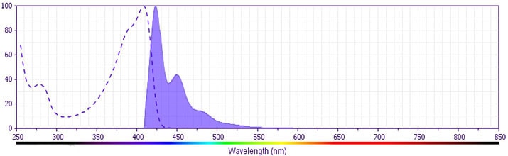
The antibody was conjugated to BD Horizon BV421 which is part of the BD Horizon Brilliant™ Violet family of dyes. With an Ex Max of 407-nm and Em Max at 421-nm, BD Horizon BV421 can be excited by the violet laser and detected in the standard Pacific Blue™ filter set (eg, 450/50-nm filter). BD Horizon BV421 conjugates are very bright, often exhibiting a 10 fold improvement in brightness compared to Pacific Blue conjugates.
Please refer to Support Documents for Quality Certificates
Global - Refer to manufacturer's instructions for use and related User Manuals and Technical data sheets before using this products as described
Comparisons, where applicable, are made against older BD Technology, manual methods or are general performance claims. Comparisons are not made against non-BD technologies, unless otherwise noted.
For Research Use Only. Not for use in diagnostic or therapeutic procedures.