Based on customer feedback, we have begun converting some of our popular BD OptiBuild™ On-Demand Reagents to our standard, off-the-shelf reagents. While converted products will have new catalog numbers, they will be available at improved pricing on select items and in familiar 50-μg unit sizes.
What you can expect:
- Off-the-shelf availability with improved pricing on select items
- Access to single lot material
- Representative data provided on the Technical Data Sheet (TDS)
- A transition period of at least 6 months for comparison testing and study completion prior to the obsolescence of BD OptiBuild™ Reagent catalog numbers
View product list highlighting the legacy BD OptiBuild™ Reagents along with the new off-the-shelf catalog numbers below.
Performance Data
Comparison of legacy BD OptiBuild™ On-Demand Reagents and converted equivalent
off-the-shelf BD Horizon™ Reagents demonstrating similar performance.
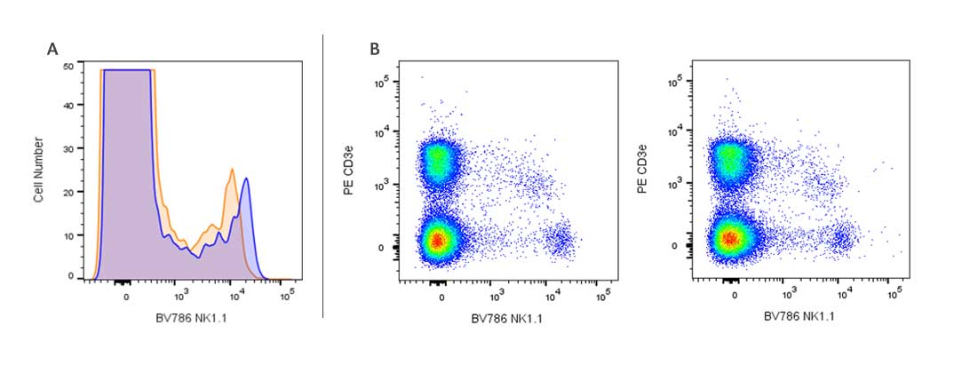
A. Flow cytometric comparison of NK-1.1 staining on mouse splenocytes using BD Horizon™ BV786 (blue histogram) or BD Optibuild™ BV786 Reagents (orange histogram) Mouse Anti-Mouse NK-1.1 antibody.
Splenic leucocytes from a C57BL/6 mouse were preincubated with Purified Rat Anti-Mouse CD16/CD32 antibody (Mouse BD Fc Block™). The cells were then stained with BV786 Mouse Anti-Mouse NK-1.1 antibodies at 0.5 ug/T. BD Via-Probe™ Cell Viability 7-AAD Solution was added to cells right before analysis. The fluorescence histograms showing NK-1.1 expression were derived from gated events with the forward and side light-scatter characteristics of viable (7-AAD-negative) splenic leucocytes.
B. Two-color flow cytometric comparison of NK-1.1 staining on mouse splenocytes using BD Horizon™ BV786 (left) or BD OptiBuild™ BV786 (right) Mouse Anti-Mouse NK-1.1 Reagents
Splenic leucocytes from a C57BL/6 mouse were preincubated with Purified Rat Anti-Mouse CD16/CD32 antibody (Mouse BD Fc Block™). The cells were then stained with PE Hamster Anti-Mouse CD3e antibody and BV786 Mouse Anti-Mouse NK-1.1 antibodies at 0.5 ug/T. BD Via-Probe™ Cell Viability 7-AAD Solution was added to cells right before analysis. Two-color flow cytometric dot plots show the correlated expression patterns of NK1.1 versus CD3e for gated events with the forward and side light-scatter characteristics of viable (7-AAD-negative) splenic leucocytes.
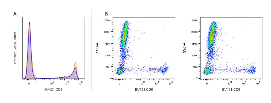
A. Flow cytometric comparison of CD8 staining on human peripheral blood lymphocytes using BD Horizon™ BV421 (blue histogram) or BD OptiBuild™ BV421 (orange histogram) Mouse Anti-Human CD8 Reagents.
Whole blood was stained with BV421 Mouse Anti-Human CD8 antibodies at 0.25 µg/test. Erythrocytes were lysed with BD FACS™ Lysing Solution. The fluorescence histogram showing CD8 expression was derived from gated events with the forward and side light-scatter characteristics of intact lymphocytes.
B. Flow cytometric comparison of CD8 staining using BD Horizon™ BV421 (left) or BD OptiBuild™ BV421 (right) Mouse Anti-Human CD8 antibody (clone SK1).
Whole blood was stained with BV421 CD8 antibodies at 0.25 µg/test. Erythrocytes were lysed with BD FACS™ Lysing Solution. The bivariate pseudocolor density plots showing the correlated expression of CD8 versus side light-scatter (SSC-A) signals were derived from gated events with the forward and side light-scatter characteristics of intact leucocyte populations.
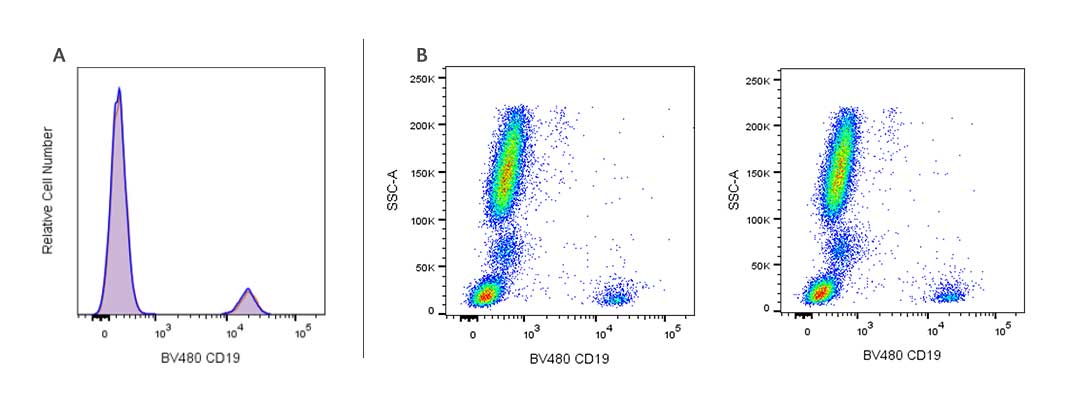
A. Flow cytometric comparison of CD19 staining on human peripheral blood lymphocytes using BD Horizon™ BV480 (blue histogram) or BD OptiBuild™ BV480 (orange histogram) Mouse Anti-Human CD19 Reagents.
Whole blood was stained with BV480 Mouse Anti-Human CD19 antibodies at 0.25 µg/test. Erythrocytes were lysed with BD FACS™ Lysing Solution. The fluorescence histograms showing CD19 expression were derived from gated events with the forward and side light-scatter characteristics of intact lymphocytes.
B. Flow cytometric comparison of CD19 staining using BD Horizon™ BV480 (left) or BD Optibuild™ BV480 (right) Mouse Anti-Human CD19 Reagents.
Whole blood was stained with BV480 Mouse Anti-Human CD19 antibodies at 0.25 µg/test. Erythrocytes were lysed with BD FACS™ Lysing Solution. The bivariate pseudocolor density plots showing the correlated expression of CD19 versus side light-scatter (SSC-A) signals were derived from gated events with the forward and side light-scatter characteristics of intact leucocyte populations.
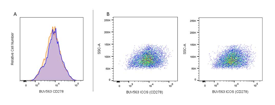
A. Flow cytometric comparison of ICOS (CD278) staining on stimulated human peripheral blood lymphocytes using BD Horizon™ BUV563 (blue histogram) or BD OptiBuild™ BUV563 (orange histogram) Mouse Anti-Human ICOS (CD278) Reagents.
Phytohemagglutinin (PHA)-stimulated (3 days) peripheral blood mononuclear cells were stained with Mouse Anti-Human ICOS (CD278) antibodies at 0.25 ug/T. BD Via-Probe™ Cell Viability 7-AAD Solution was added to cells right before analysis. The fluorescence histogram showing ICOS (CD278) expression was derived from gated events with the forward and side light-scatter characteristics of viable (7-AAD-negative) lymphocytes.
B. Flow cytometric comparison of ICOS (CD278) staining on stimulated human peripheral blood lymphocytes using BD Horizon™ BUV563 (left) or BD Optibuild™ BUV563 (right) Mouse Anti-Human ICOS (CD278) antibody.
Phytohemagglutinin (PHA)-stimulated (3 days) peripheral blood mononuclear cells were stained with Mouse Anti-Human ICOS (CD278) antibodies at 0.25 ug/T. BD Via-Probe™ Cell Viability 7-AAD Solution was added to cells right before analysis. The bivariate pseudocolor density plots showing the correlated expression of ICOS (CD278) versus side light-scatter (SSC-A) signals were derived from gated events with the forward and side light-scatter characteristics of viable (7-AAD-negative) lymphocytes.
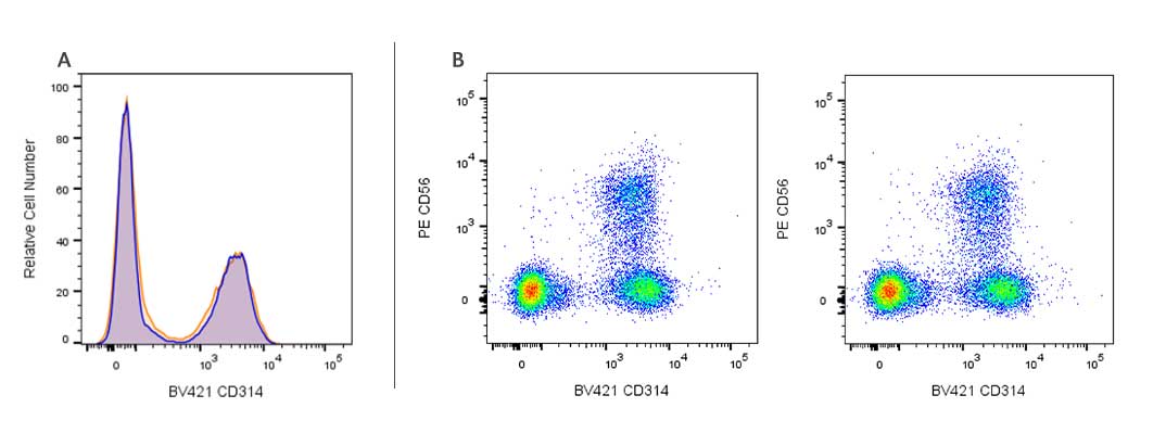
A. Flow cytometric comparison of CD314 (NKG2D) staining on human peripheral blood lymphocytes using BD Horizon™ BV421 (blue histogram) or BD OptiBuild™ BV421 (orange histogram) Mouse Anti-Human CD314 (NKG2D) Reagents.
Whole blood was stained with Mouse Anti-Human CD314 (NKG2D) at 0.5 µg/test. Erythrocytes were lysed with BD FACS™ Lysing Solution. The fluorescence histogram showing CD314 (NKG2D) expression was derived from gated events with the forward and side light-scatter characteristics of intact lymphocytes.
B. Two-color flow cytometric comparison of CD314 (NKG2D) staining on human peripheral blood lymphocytes using BD Horizon™ BV421 (A) or BD OptiBuild™ BV421 (B) Mouse Anti-Human CD314 (NKG2D) Reagents.
Whole blood was stained with PE Anti-Human CD56 and BV421 Mouse Anti-Human CD314 (NKG2D) antibodies at 0.5 µg/test. Erythrocytes were lysed with BD FACS™ Lysing Solution. A bivariate pseudocolor density plot showing the correlated expression of CD314 (NKG2D) versus CD56 was derived from gated events with the forward and side light-scatter characteristics of intact lymphocytes.
Product Transition Chart
| Legacy BD OptiBuild™ Reagent Catalog Number | Species | Description | Clone | Isotype | Format | New BD Horizon™ Reagent Catalog Number |
|---|---|---|---|---|---|---|
| 740076 | Human | Hu CD56 BV421 B159 50ug | B159 | Mouse IgG1, κ | BV421 | 568219 |
| 740082 | Human | Hu HLA-A2 BV421 BB7.2 50ug | BB7.2 | Mouse IgG2b, κ | BV421 | 568757 |
| 740083 | Human | Hu CD45RA BV421 5H9 50ug | 5H9 | Mouse IgG1, κ | BV421 | 569620 |
| 740093 | Human | Hu CD8 BV421 SK1 50ug | SK1 | Mouse IgG1, κ | BV421 | 568217 |
| 740101 | Human | Hu CD20 BV510 L27 50ug | L27 | Mouse IgG1, κ | BV510 | 569502 |
| 740157 | Mouse | Ms Ly-6G BV510 1A8 50ug | 1A8 | Rat (LEW) IgG2a, κ | BV510 | 569685 |
| 740161 | Human | Hu CD4 BV510 RPA-T4 50ug | RPA-T4 | Mouse IgG1, κ | BV510 | 569388 |
| 740176 | Human | Hu CD95 BV510 DX2 50ug | DX2 | Mouse (C3H) IgG1, κ | BV510 | 569687 |
| 740180 | Human | Hu CD206 BV510 19.2 50ug | 19.2 | Mouse IgG1, κ | BV510 | 569618 |
| 740187 | Human | Hu CD3 BV510 SP34-2 50ug | SP34-2 | Mouse IgG1, κ | BV510 | 569486 |
| 740202 | Human | Hu CD3 BV510 SK7 50ug | SK7 | Mouse IgG1, κ | BV510 | 569250 |
| 740208 | Mouse | Ms CD4 BUV395 RM4-5 50ug | RM4-5 | Rat IgG2a, κ | BUV395 | 568375 |
| 740215 | Mouse | Ms CD44 BUV395 IM7 50ug | IM7 | Rat IgG2b, κ | BUV395 | 568507 |
| 740218 | Mouse | Ms CD62L BUV395 MEL-14 50ug | MEL-14 | Rat IgG2a, κ | BUV395 | 569400 |
| 740220 | Mouse | Ms CD69 BUV395 H1.2F3 50ug | H1.2F3 | Ham IgG2, λ3 | BUV395 | 569367 |
| 740238 | Mouse | Ms CD103 BUV395 M290 50ug | M290 | Rat IgG2a, κ | BUV395 | 568715 |
| 740267 | Human | Hu CCR7 (CD197) BUV395 3D12 50ug | 3D12 | Rat IgG2a, κ | BUV395 | 568681 |
| 740268 | Mouse | Ms CD3 BUV395 17A2 50ug | 17A2 | Rat IgG2b, κ | BUV395 | 569614 |
| 740286 | Human | Hu CD14 BUV395 M5E2 50ug | M5E2 | Mouse IgG2a, κ | BUV395 | 569102 |
| 740287 | Human | Hu CD19 BUV395 HIB19 50ug | HIB19 | Mouse IgG1, κ | BUV395 | 569107 |
| 740290 | Human | Hu CD25 (IL-2 RCPTR α) BUV395 M-A251 50ug | M-A251 | Mouse IgG1, κ | BUV395 | 569353 |
| 740291 | Human | Hu CD27 BUV395 M-T271 50ug | M-T271 | Mouse (BALB/c) IgG1 κ | BUV395 | 569712 |
| 740293 | Human | Hu CD33 BUV395 WM53 50ug | WM53 | Mouse IgG1, κ | BUV395 | 568374 |
| 740298 | Human | Hu CD45RA BUV395 HI100 50ug | HI100 | Mouse IgG2b, κ | BUV395 | 568712 |
| 740303 | Human | Hu CD8 BUV395 HIT8A 50ug | HIT8a | Mouse IgG1, κ | BUV395 | 569178 |
| 740308 | Human | Hu CD28 BUV395 CD28.2 50ug | CD28.2 | Mouse IgG1, κ | BUV395 | 569160 |
| 740315 | Human | Hu CD45RA BUV395 5H9 50ug | 5H9 | Mouse IgG1, κ | BUV395 | 569489 |
| 740326 | Mouse | Ms CD335 (NKp46) BUV395 29A1.4 50ug | 29A1.4 | Rat IgG2a, κ | BUV395 | 568630 |
| 740333 | Human | Hu CD20 BV605 L27 50ug | L27 | Mouse IgG1, κ | BV605 | 569503 |
| 740363 | Mouse | Ms CD49b BV605 HMa2 50ug | HMα2 | Ham IgG1, κ | BV605 | 569508 |
| 740379 | Human | HU CXCR5 (CD185) BV605 RF8B2 50ug | RF8B2 | Rat IgG2b, κ | BV605 | 569174 |
| 740391 | Human | Hu CD2 BV605 RPA-2.10 50ug | RPA-2.10 | Mouse IgG1, κ | BV605 | 569168 |
| 740392 | Human | Hu CD7 BV605 M-T701 50ug | M-T701 | Mouse IgG1, κ | BV605 | 569104 |
| 740394 | Human | Hu CD19 BV605 HIB19 50ug | HIB19 | Mouse IgG1, κ | BV605 | 569363 |
| 740397 | Human | Hu CD25 BV605 M-A251 50ug | M-A251 | Mouse IgG1, κ | BV605 | 569171 |
| 740398 | Human | Hu CD27 BV605 M-T271 50ug | M-T271 | Mouse IgG1, κ | BV605 | 569170 |
| 740401 | Human | Hu CD38 BV605 HIT2 50ug | HIT2 | Mouse IgG1 κ | BV605 | 569699 |
| 740406 | Human | Hu CD64 BV605 10.1 50ug | 10.1 | Mouse IgG1, κ | BV605 | 569172 |
| 740407 | Human | Hu HLA-ABC BV605 G46-2.6 50ug | G46-2.6 | Mouse IgG1, κ | BV605 | 569512 |
| 740411 | Human | Hu CD8 BV605 HIT8A 50ug | HIT8a | Mouse IgG1, κ | BV605 | 569169 |
| 740417 | Human | Hu CD206 BV605 19.2 50ug | 19.2 | Mouse IgG1, κ | BV605 | 569177 |
| 740421 | Human | Hu CD141 BV605 1A4 50ug | 1A4 | Mouse (BALB/c) IgG1 κ | BV605 | 569684 |
| 740426 | Human | Hu CD274 BV605 MIH1 50ug | MIH1 | Mouse IgG1, κ | BV605 | 569469 |
| 740436 | Human | Hu CD16 BV605 B73.1 50ug | B73.1 | Mouse IgG1, κ | BV605 | 569175 |
| 740455 | Mouse | Ms CD44 BV650 IM7 50ug | IM7 | Rat IgG2b κ | BV650 | 569707 |
| 740460 | Mouse | Ms CD69 BV650 H1.2F3 50ug | H1.2F3 | Armenian Hamster IgG1 λ3 | BV650 | 569688 |
| 740528 | Human | Hu CXCR5 (CD185) BV650 RF8B2 50ug | RF8B2 | Rat IgG2b, κ | BV650 | 569547 |
| 740530 | Mouse | Ms CD3 BV650 17A2 50ug | 17A2 | Rat (SD) IgG2b κ | BV650 | 569683 |
| 740553 | Mouse | Ms KLRG1 BV650 2F1 50ug | 2F1 | Ham IgG2, κ | BV650 | 569548 |
| 740566 | Human | Hu CD11b BV650 ICRF44 50ug | ICRF44 | Mouse IgG1 κ | BV650 | 569704 |
| 740574 | Human | Hu CD38 BV650 HIT2 50ug | HIT2 | Mouse IgG1, κ | BV650 | 569391 |
| 740581 | Human | Hu HLA-ABC BV650 G46-2.6 50ug | G46-2.6 | Mouse IgG1, κ | BV650 | 569393 |
| 740604 | Human | Hu CD141 BV650 1A4 50ug | 1A4 | Mouse IgG1, κ | BV650 | 569392 |
| 740610 | NHP | NHP CD45 BV650 D058-1283 50 ug | D058-1283 | Mouse IgG1, κ | BV650 | 569619 |
| 740627 | Mouse | Ms CD335 (NKp46) BV650 29A1.4 50ug | 29A1.4 | Rat IgG2a, κ | BV650 | 569494 |
| 740660 | Mouse | Ms CD62L BV711 MEL-14 50ug | MEL-14 | Rat IgG2a, κ | BV711 | 568286 |
| 740663 | Mouse | Ms NK-1.1 BV711 PK136 50ug | PK136 | Mouse (C3H x BALB/c) IgG2a κ | BV711 | 569723 |
| 740664 | Mouse | Ms CD69 BV711 H1.2F3 50ug | H1.2F3 | Armenian Hamster IgG1 λ3 | BV711 | 569691 |
| 740722 | Mouse | Hu CD123 BV711 7G3 50ug | 7G3 | Mouse IgG2a, κ | BV711 | 568228 |
| 740769 | Human | Hu CD4 BV711 RPA-T4 50ug | RPA-T4 | Mouse IgG1, κ | BV711 | 568371 |
| 740771 | Human | Hu CD11b BV711 ICRF44 50ug | ICRF44 | Mouse IgG1, κ | BV711 | 568229 |
| 740782 | Human | Hu CD64 BV711 10.1 50ug | 10.1 | Mouse (BALB/c) IgG1 κ | BV711 | 569686 |
| 740798 | Human | Hu CD137 BV711 4B4-1 50ug | 4B4-1 | Mouse IgG1 κ | BV711 | 569689 |
| 740801 | Human | Hu CD80 BV711 L307.4 50ug | L307.4 | Mouse IgG1, κ | BV711 | 568227 |
| 740832 | Human | Hu CD3 BV711 SK7 50ug | SK7 | Mouse (BALB/c) IgG1 κ | BV711 | 569787 |
| 740853 | Mouse | Ms NK-1.1 BV786 PK136 50ug | PK136 | Mouse IgG2a | BV786 | 568224 |
| 740861 | Mouse | CD11B BV786 M1/70 50ug | M1/70 | Rat IgG2b, κ | BV786 | 569504 |
| 740880 | Mouse | Ms CD138 BV786 281-2 50ug | 281-2 | Rat (F344) IgG2a, κ | BV786 | 569692 |
| 740889 | Mouse | Ms CD45.1 BV786 A20 50ug | A20 | Mouse A.SW IgG2a, κ | BV786 | 569696 |
| 740953 | Mouse | Ms Ly-6G BV786 1A8 50ug | 1A8 | Rat IgG2a, κ | BV786 | 569406 |
| 740962 | Human | Hu CD4 BV786 RPA-T4 50ug | RPA-T4 | Mouse IgG1, κ | BV786 | 568368 |
| 740965 | Human | Hu CD11b BV786 ICRF44 50ug | ICRF44 | Mouse IgG1, κ | BV786 | 569703 |
| 740966 | Human | Hu CD11c BV786 B-ly6 50ug | B-ly6 | Mouse IgG1, κ | BV786 | 568220 |
| 740968 | Human | Hu CD19 BV786 HIB19 50ug | HIB19 | Mouse IgG1 κ | BV786 | 569698 |
| 740971 | Human | Hu CD24 BV786 ML5 50ug | ML5 | Mouse IgG2a | BV786 | 568225 |
| 740974 | Human | Hu CD33 BV786 WM53 50ug | WM53 | Mouse (BALB/c) IgG1, κ | BV786 | 569790 |
| 740981 | Human | Hu CD62L BV786 DREG-56 50ug | DREG-56 | Mouse IgG1 κ | BV786 | 569695 |
| 740991 | Human | Hu CD95 BV786 DX2 50ug | DX2 | Mouse IgG1, κ | BV786 | 568505 |
| 740992 | Human | Hu CD81 (TAPA-1) BV786 JS-81 50ug | JS-81 | Mouse IgG1, κ | BV786 | 568522 |
| 740996 | Human | Hu CD28 BV786 CD28.2 50ug | CD28.2 | Mouse (C3H x BALB/c) IgG1, κ | BV786 | 569697 |
| 741003 | Human | Hu CD163 BV786 GHI/61 50ug | GHI/61 | Mouse IgG1, κ | BV786 | 568221 |
| 741006 | Human | Hu CD141 BV786 1A4 50ug | 1A4 | Mouse IgG1, κ | BV786 | 568218 |
| 741013 | Human | Hu CD15 BV786 W6D3 50ug | W6D3 | Mouse IgG1, κ | BV786 | 568288 |
| 741024 | Mouse | Ms CD64 A/B ALOATG BV786 X54-5/7.1 50ug | X54-5/7.1 | Mouse IgG1, κ | BV786 | 569507 |
| 741029 | Mouse | Ms CD335 (NKp46) BV786 29A1.4 50ug | 29A1.4 | Rat IgG2a κ | BV786 | 569694 |
| 741050 | Mouse | Ms CD4 BUV496 RM4-5 50ug | RM4-5 | Rat IgG2a, κ | BUV496 | 569180 |
| 741057 | Mouse | Ms CD44 BUV496 IM7 50ug | IM7 | Rat IgG2b κ | BUV496 | 569706 |
| 741062 | Mouse | Ms NK-1.1 BUV496 PK136 50ug | PK136 | Mouse C3H x BALB/c IgG2a, κ | BUV496 | 569716 |
| 741092 | Mouse | Ms CD45.2 BUV496 104 50ug | 104 | Mouse (SJL) IgG2a, κ | BUV496 | 569670 |
| 741117 | Mouse | Ms CD3 BUV496 17A2 50ug | 17A2 | Rat (SD) IgG2b κ | BUV496 | 569671 |
| 741134 | Human | Hu CD4 BUV496 RPA-T4 50ug | RPA-T4 | Mouse IgG1, κ | BUV496 | 569179 |
| 741138 | Human | Hu CD11b BUV496 ICRF44 50ug | ICRF44 | Mouse IgG1 κ | BUV496 | 569702 |
| 741206 | Human | Hu CD3 BUV496 SK7 50ug | SK7 | Mouse (BALB/c) IgG1 κ | BUV496 | 569786 |
| 741217 | Mouse | Ms CD4 BUV563 RM4-5 50ug | RM4-5 | Rat IgG2a, κ | BUV563 | 569182 |
| 741227 | Mouse | Ms CD44 BUV563 IM7 50ug | IM7 | Rat IgG2b κ | BUV563 | 569705 |
| 741242 | Mouse, Human | CD11b BUV563 M1/70 50ug | M1/70 | Rat (DA) IgG2b κ | BUV563 | 569711 |
| 741357 | Human | Hu CD11b BUV563 ICRF44 50ug | ICRF44 | Mouse IgG1 κ | BUV563 | 569701 |
| 741361 | Human | Hu CD19 BUV563 HIB19 50ug | HIB19 | Mouse IgG1 κ | BUV563 | 569676 |
| 741421 | Human | Hu ICOS (CD278) BUV563 DX29 50ug | DX29 | Mouse IgG1, κ | BUV563A | 568215 |
| 741448 | Human | Hu CD3 BUV563 SK7 50ug | SK7 | Mouse (BALB/c) IgG1, κ | BUV563 | 569785 |
| 741461 | Human | Ms CD4 BUV661 RM4-5 50ug | RM4-5 | Rat (DA) IgG2a, κ | BUV661 | 569752 |
| 741597 | Human | Hu CD4 BUV661 RPA-T4 50ug | RPA-T4 | Mouse IgG1 κ | BUV661 | 569782 |
| 741642 | Human | Hu CD137 BUV661 4B4-1 50ug | 4B4-1 | Mouse IgG1 κ | BUV661 | 569682 |
| 741692 | Human | Hu CD3 BUV661 SK7 50ug | SK7 | Mouse (BALB/c) IgG1, κ | BUV661 | 569784 |
| 741813 | Mouse | Ms Ly-6G BUV737 1A8 50ug | 1A8 | Rat IgG2a, κ | BUV737 | 568346 |
| 741823 | Human | Hu CD4 BUV737 RPA-T4 50ug | RPA-T4 | Mouse IgG1, κ | BUV737 | 568369 |
| 741826 | Human | Hu CD11b BUV737 ICRF44 50ug | ICRF44 | Mouse IgG1, κ | BUV737 | 568332 |
| 741827 | Human | Hu CD11c BV737 B-ly6 50ug | B-ly6 | Mouse IgG1, κ | BUV737 | 568328 |
| 741842 | Human | Hu CD56 (NCAM-1) BUV737 B159 50ug | B159 | Mouse IgG1, κ | BUV737 | 569191 |
| 741843 | Human | Hu CD62L BUV737 DREG-56 50ug | DREG-56 | Mouse IgG1, κ | BUV737 | 568329 |
| 741861 | Human | Hu CD137 BUV737 4B4-1 50ug | 4B4-1 | Mouse IgG1, κ | BUV737 | 568348 |
| 741865 | Human | Hu CD80 BUV737 L307.4 50ug | L307.4 | Mouse IgG1, κ | BUV737 | 568364 |
| 741872 | Human | Hu CD3 BUV737 SP34-2 50ug | SP34-2 | Mouse IgG1, λ1 | BUV737 | 568353 |
| 741877 | Mouse | Ms CD274 BUV737 MIH5 50ug | MIH5 | Rat IgG2a, λ | BUV737 | 568361 |
| 741912 | Mouse | Ms CD4 BUV805 RM4-5 50ug | RM4-5 | Rat IgG2a, κ | BUV805 | 569193 |
| 741924 | Mouse | Ms CD62L BUV805 MEL-14 50ug | MEL-14 | Rat IgG2a, κ | BUV563 | 569201 |
| 741926 | Mouse | Ms NK-1.1 BUV805 PK136 50ug | PK136 | Mouse IgG2a, κ | BUV805 | 569164 |
| 741934 | Ms/Hu | CD11b BUV805 M1/70 50ug | M1/70 | Rat IgG2b, κ | BUV805 | 568345 |
| 741957 | Mouse | Ms CD45.2 BUV805 104 50ug | 104 | Mouse IgG2a, κ | BUV805 | 569200 |
| 741958 | Mouse | Ms CD45.1 BUV805 A20 50ug | A20 | Mouse (A.SW) IgG2a, κ | BUV805 | 569681 |
| 741982 | Mouse | Ms CD3 BUV805 17A2 50ug | 17A2 | Rat IgG2b, κ | BUV805 | 569192 |
| 742000 | Human | Hu CD4 BUV805 RPA-T4 50ug | RPA-T4 | Mouse IgG1, κ | BUV563 | 569196 |
| 742004 | Human | Hu CD11b BUV805 ICRF44 50ug | ICRF44 | Mouse IgG1, κ | BUV805 | 569700 |
| 742007 | Human | Hu CD19 BUV805 HIB19 50ug | HIB19 | Mouse IgG1, κ | BUV805 | 568331 |
| 742020 | Human | Hu CD45RA BUV805 HI100 50ug | HI100 | Mouse IgG2b, κ | BUV805 | 568330 |
| 742024 | Human | Hu CD62L BUV805 DREG-56 50ug | DREG-56 | Mouse IgG1, κ | BUV805 | 569161 |
| 742053 | Human | Hu CD3 BUV805 SP34-2 50ug | SP34-2 | Mouse IgG1, λ1 | BUV805 | 568354 |
| 742069 | Human | Hu CD14 BUV805 MphiP9 50ug | MphiP9 | Mouse IgG2b, κ | BUV805 | 568333 |
| 742075 | Human | Hu CD16 (FcγRIII) BUV805 B73.1 50ug | B73.1 | Mouse IgG1, κ | BUV805 | 569166 |
| 742217 | Human | Hu CD33 BB700 WM53 50ug | WM53 | Mouse IgG1, κ | BB700 | 568283 |
| 742229 | Human | Hu CD8 BB700 HIT8a 50ug | HIT8a | Mouse IgG1, κ | BB700 | 568284 |
| 742390 | Human | Hu CD8b BV421 2ST8.5H7 50ug | 2ST8.5H7 | Mouse IgG2a, κ | BV421 | 568373 |
| 742393 | Human | Hu CD8b BV650 2ST8.5H7 50ug | 2ST8.5H7 | Mouse IgG2a, κ | BV650 | 569550 |
| 742484 | Mouse | Ms TCR b Chain BV786 H57-597 50ug | H57-597 | Ham IgG2, λ1 | BV786 | 568222 |
| 742485 | Mouse | Ms TCR Beta Chain BUV395 H57-597 50ug | H57-597 | Ham IgG2, λ1 | BUV395 | 569248 |
| 742550 | Human | Hu CD5 BV605 L17F12 50ug | L17F12 | Mouse IgG2a, κ | BV605 | 569239 |
| 742623 | Human | Hu CD3 BV605 UCHT1 50ug | UCHT1 | Mouse IgG1, κ | BV605 | 569100 |
| 742731 | Human | Hu CD27 BV421 L128 50ug | L128 | Mouse IgG1, κ | BV421 | 568226 |
| 742749 | Human | Hu CD1c BV650 F10/21A3 50ug | F10/21A3 | Mouse IgG1, κ | BV650 | 569617 |
| 742856 | Human | Hu TIM-3 (CD366) BV605 7D3 50ug | 7D3 | Mouse IgG1, κ | BV605 | 569245 |
| 742857 | Human | Hu TIM-3 (CD366) BV786 7D3 50ug | 7D3 | Mouse IgG1, κ | BV786 | 568504 |
| 743155 | Mouse | Ms CD4 BV510 GK1.5 50ug | GK1.5 | Rat IgG2b, κ | BV510 | 569249 |
| 743172 | Human | Hu CD337 (NKp30) BV786 P30-15 50ug | P30-15 | Mouse IgG1, κ | BV786 | 568223 |
| 743280 | Mouse | Ms F4/80 BV510 T45-2342 50ug | T45-2342 | Rat IgG2a, κ | BV510 | 569615 |
| 743281 | Mouse | Ms F4/80 BV605 T45-2342 50ug | T45-2342 | Rat IgG2a, κ | BV605 | 569237 |
| 743283 | Human | Hu CD134 BV421 ACT35 50ug | ACT35 | Mouse IgG1, κ | BV421 | 568213 |
| 743307 | Human | Hu CD71 BV650 M-A712 50ug | M-A712 | Mouse IgG2a, κ | BV650 | 569511 |
| 743308 | Human | Hu CD71 BUV395 M-A712 50ug | M-A712 | Mouse IgG2a, κ | BUV395 | 568523 |
| 743558 | Human | Hu CD314 (NKG2D) BV421 1D11 50ug | 1D11 | Mouse IgG1, κ | BV421 | 568106 |
| 743611 | Human | Hu CD20 BV786 2H7 50ug | 2H7 | Mouse IgG2b, κ | BV786 | 568713 |
| 743652 | Human | Hu CD103 BV605 BER-ACT8 50ug | Ber-ACT8 | Mouse IgG1, κ | BV605 | 569162 |
| 743654 | Human | Hu CD103 BV786 Ber-ACT8 50ug | Ber-ACT8 | Mouse IgG1, κ | BV786 | 568274 |
| 743876 | Mouse | Ms I-A, I-E BUV395 2G9 50ug | 2G9 | Rat IgG2a, κ | BUV395 | 569244 |
| 744023 | Human | Hu CD1a BV605 HI149 50ug | HI149 | Mouse IgG1, κ | BV605 | 569690 |
| 744436 | Human | Hu CD11c BV605 S-HCL-3 50ug | S-HCL-3 | Mouse IgG2b, κ | BV605 | 569488 |
| 744471 | Mouse | Ms CD24 BUV395 M1/69 50ug | M1/69 | Rat (DA) IgG2b κ | BUV395 | 569669 |
| 744546 | Mouse | Ms CD279 (PD-1) BV650 J43 50ug | J43 | Ham IgG2, κ | BV650 | 569506 |
| 744547 | Mouse | Ms CD279 (PD-1) BV711 J43 50ug | J43 | Ham IgG2, κ | BV711 | 568298 |
| 744904 | Human | Hu CD34 BV421 8G12 50ug | 8G12 | Mouse IgG1, κ | BV421 | 568758 |
| 744930 | Human | Hu CD8b BV650 2ST8.5H7 50ug | DX29 | Mouse IgG1, κ | BV510 | 569549 |
| 744985 | Human | Hu LAG-3 (CD223) BV510 T47-530 50ug | T47-530 | Mouse IgG1, κ | BV510 | 569616 |
| 745091 | Human | Hu CD163 BV605 GHI/61 50ug | GHI/61 | Mouse IgG1, κ | BV605 | 569202 |
| 745100 | Human | Hu ICOS (CD278) BV605 DX29 50ug | DX29 | Mouse IgG1, κ | BV605 | 569173 |
| 745105 | Human | Hu CD34 BV605 563 50ug | 563 | Mouse IgG1, κ | BV605 | 569594 |
| 745191 | Human | Hu CD13 BV605 L138 50ug | L138 | Mouse IgG1, κ | BV605 | 569238 |
| 745205 | Human | Hu CD2 BV605 S5.2 50ug | S5.2 | Mouse IgG2a | BV605 | 569106 |
| 745359 | Human | Hu Gma Dta TCR BV650 11F2 50ug | 11F2 | Mouse IgG1 | BV650 | 569510 |
| 745505 | Human | Hu Gma Dta TCR BV711 11F2 50ug | 11F2 | Mouse IgG1 | BV711 | 568490 |
| 745640 | Human | Hu LAG-3 (CD223) BUV395 T47-530 50ug | T47-530 | Mouse IgG1, κ | BUV395 | 569247 |
| 745733 | Human | Hu CD117 (c-Kit) BUV395 104D2 50ug | 104D2 | Mouse IgG1 | BUV395 | 568489 |
| 745787 | Human | Hu CD138 BB700 MI15 50ug | MI15 | Mouse IgG1, κ | BB700 | 568282 |
| 745790 | Human | Hu CD14 BB700 M5E2 50ug | M5E2 | Mouse IgG2a, κ | BB700 | 568525 |
| 745981 | Human | Hu CD4 BB700 RPA-T4 50ug | RPA-T4 | Mouse IgG1, κ | BB700 | 568370 |
| 746313 | Human | Hu CD3 BV480 HIT3A 50ug | HIT3a | Mouse IgG2a, κ | BV480 | 568216 |
| 746457 | Human | Hu CD19 BV480 HIB19 50ug | HIB19 | Mouse IgG1, κ | BV480 | 568214 |
| 746541 | Human | Hu CD4 BV480 RPA-T4 50ug | RPA-T4 | Mouse IgG1, κ | BV480 | 569781 |
| 747353 | Human | Hu CD137 BV750 4B4-1 50ug | 4B4-1 | Mouse (BALB/c) IgG1, κ | BV750 | 569693 |
| 747520 | Human | Hu CD69 BB700 FN50 50ug | FN50 (also known as FN 50) | Mouse IgG1, κ | BB700 | 569399 |
| 747522 | Human | Hu CD69 BV750 FN50 50ug | FN50 | Mouse IgG1, κ | BV750 | 568285 |
| 747750 | Human | Hu CD90 BV605 5E10 50ug | 5E10 | Mouse IgG1, κ | BV605 | 569401 |
| 748267 | Mouse | Ms CD279 (PD-1) BV605 RMP1-30 50ug | RMP1-30 | Rat IgG2b, κ | BV605 | 569105 |
| 748268 | Mouse | Ms CD279 (PD-1) BV421 RMP1-30 50ug | RMP1-30 | Rat (SD) IgG2b κ | BV421 | 569780 |
| 748338 | Human | Hu HLA-DR BUV805 G46-6 50ug | G46-6 | Mouse IgG2a, κ | BUV805 | 568335 |
| 748339 | Human | Hu HLA-DR BUV737 G46-6 50ug | G46-6 | Mouse IgG2a, κ | BUV737 | 568351 |
| 748370 | Mouse | Ms CD45 BUV805 30-F11 50ug | 30-F11 | Rat IgG2b, κ | BUV805 | 568336 |
| 748371 | Mouse | Ms CD45 BUV737 30-F11 50ug | 30-F11 | Rat IgG2b, κ | BUV737 | 568344 |
| 748502 | Human | Hu CD103 BUV737 Ber-ACT8 50ug | Ber-ACT8 | Mouse IgG1, κ | BUV737 | 568350 |
| 748535 | Mouse | Ms CD8a BUV563 53-6.7 50ug | 53-6.7 | Rat IgG2a, κ | BUV563 | 569185 |
| 748569 | Human | Hu CD3 BUV563 UCHT1 50ug | UCHT1 | Mouse BALB/c IgG1, κ | BUV563 | 569789 |
| 748609 | Human | Hu CD56 BUV737 MY31 50ug | MY31 | Mouse (BALB/c X C57BL/6) IgG1, κ | BUV737 | 569722 |
| 748704 | Human | Hu CD27 BUV805 L128 50ug | L128 | Mouse IgG1, κ | BUV805 | 569167 |
| 748719 | Human | Hu CD45 BUV737 HI30 50ug | HI30 | Mouse IgG1, κ | BUV737 | 568524 |
| 748720 | Human | Hu CD45 BUV563 HI30 50 ug | HI30 | Mouse IgG1, κ | BUV563 | 569675 |
| 748763 | Human | Hu CD69 (Leu-23) BUV805 FN50 50ug | FN50 | Mouse IgG1, κ | BUV805 | 569163 |
| 748820 | Human | Hu TIM-3 (CD366) BUV737 7D3 50ug | 7D3 | Mouse IgG1, κ | BUV737 | 568680 |
| 748845 | Mouse | Ms I-A, I-E BUV737 M5/114.15.2 50ug | M5/114.15.2 | Rat IgG2b, κ | BUV737 | 569176 |
| 748850 | Human | Hu CD16 BUV805 3G8 50ug | 3G8 | Mouse IgG1, κ | BUV805 | 569165 |
| 748851 | Human | Hu CD16 BUV563 3G8 50ug | 3G8 | Mouse IgG1, κ | BUV563 | 568289 |
| 748867 | Mouse | Ms CD45R/B220 BUV805 RA3-6B2 50ug | RA3-6B2 | Mouse IgG1, κ | BUV563 | 569199 |
| 749027 | Mouse | Ms CD19 BUV805 1D3 50ug | 1D3 | Mouse IgG1, κ | BUV805 | 568287 |
| 749038 | Human | Ms CD11c BUV805 N418 50ug | N418 | Armenian Hamster IgG2 | BUV805 | 569713 |
| 749039 | Mouse | Ms CD11c BUV737 N418 50ug | N418 | Ham IgG2, κ | BUV737 | 569236 |
| 749173 | Human | Hu CD19 BUV805 SJ25C1 50ug | SJ25C1 | Mouse IgG1, κ | BUV805 | 568290 |
| 749306 | Mouse | Ms CD279 (PD-1) BUV737 RMP1-30 50ug | RMP1-30 | Rat IgG2b, κ | BUV737 | 568363 |
| 749366 | Human | Hu CD8 BUV805 RPA-T8 50ug | RPA-T8 | Mouse IgG2a, κ | BUV805 | 568334 |
| 749367 | Human | Hu CD8 BUV737 RPA-T8 50ug | RPA-T8 | Mouse IgG1, κ | BUV737 | 569189 |
| 749393 | Mouse | Ms CD103 BUV737 2E7 50ug | 2E7 | Armenian Hamster IgG2, κ | BUV737 | 569678 |
| 749422 | Mouse | Ms CD279 (PD-1) BUV737 J43 50ug | J43 | Ham IgG2, κ | BUV737 | 568362 |
| 749655 | Human | Hu CCR7 (CD197) BUV395 2-L1-A 50ug | 2-L1-A | Mouse IgG1, κ | BUV395 | 569108 |
| 749864 | Human, Mouse | CD11b BUV496 M1/70 50ug | M1/70 | Rat (DA) IgG2b, κ | BUV496 | 569708 |
| 749866 | Human | Hu HLA-DR BUV496 G46-6 50ug | G46-6 | Mouse IgG2a κ | BUV496 | 569674 |
| 749889 | Mouse | Ms CD45 BUV496 30-F11 50ug | 30-F11 | Rat IgG2b κ | BUV496 | 569673 |
| 749954 | Human | Hu CD20 BUV496 2H7 50ug | 2H7 | Mouse (C57BL/6) IgG2b κ | BUV496 | 569672 |
| 749967 | Human | Hu CD39 (ENTPD1) BUV661 TU66 50ug | TU66 | Mouse IgG2b, κ | BUV661 | 569788 |
| 750023 | Mouse | Ms CD8a BUV661 53-6.7 50ug | 53-6.7 | Rat IgG2a, κ | BUV563 | 569186 |
| 750024 | Mouse | Ms CD8a BUV496 53-6.7 50ug | 53-6.7 | Rat IgG2a, κ | BUV496 | 569181 |
| 750179 | Human | Hu CD45 BUV496 HI30 50ug | HI30 | Mouse IgG1, κ | BUV496 | 569101 |
| 750260 | Human | Hu CD279 (PD-1) BUV661 EH12.1 50ug | EH12.1 | Mouse IgG1, κ | BUV661 | 569246 |
| 750591 | Human | Hu CD4 BUV496 L200 50ug | L200 | Mouse IgG1, κ | BUV496 | 569505 |
| 750696 | Human | Hu CD196 (CCR6) BUV661 11A9 50ug | 11A9 | Mouse IgG1, κ | BUV661 | 569509 |
| 750699 | Human | Hu CD8 BUV661 RPA-T8 50ug | RPA-T8 | Mouse IgG1, κ | BUV563 | 569187 |
| 750971 | Human | Hu CD3 BUV737 OKT3 50ug | OKT3 | Mouse IgG2a, κ | BUV737 | 569188 |
| 750986 | Human | Hu CD4 BV480 OKT4 50ug | OKT4 | Mouse IgG2b, κ | BV480 | 568372 |
| 751273 | Human | Hu CD19 BUV615 HIB19 50ug | HIB19 | Mouse IgG1 κ | BUV615 | 569677 |
| 751518 | Human | Hu CD8 BUV615 RPA-T8 50ug | RPA-T8 | Mouse IgG1, κ | BUV615 | 569783 |
| 751580 | Mouse | Ms CD45R/B220 BUV615 RA3-6B2 50ug | RA3-6B2 | Rat IgG2a, κ | BUV615 | 569724 |
| 751681 | Human | Hu CD27 BUV737 O323 50ug | O323 | Mouse IgG1, κ | BUV737 | 569714 |
| 751682 | Human | Hu CD27 BUV805 O323 50ug | O323 | Mouse IgG1, κ | BUV805 | 569235 |
| 751840 | Human | Hu IL-3RΑ (CD123) BUV805 6H6 50ug | 6H6 | Mouse (BALB/c) IgG1, κ | BUV805 | 569680 |
BV: BD Horizon Brilliant Violet™ Reagents
BUV: BD Horizon Brilliant™ Ultraviolet Reagents
The product list will be updated with future releases
For Research Use Only. Not for use in therapeutic or diagnostic procedures.