-
Reagents
- Flow Cytometry Reagents
-
Western Blotting and Molecular Reagents
- Immunoassay Reagents
-
Single-Cell Multiomics Reagents
- BD® OMICS-Guard Sample Preservation Buffer
- BD® AbSeq Assay
- BD® Single-Cell Multiplexing Kit
- BD Rhapsody™ ATAC-Seq Assays
- BD Rhapsody™ Whole Transcriptome Analysis (WTA) Amplification Kit
- BD Rhapsody™ TCR/BCR Next Multiomic Assays
- BD Rhapsody™ Targeted mRNA Kits
- BD Rhapsody™ Accessory Kits
- BD® OMICS-One Protein Panels
- BD OMICS-One™ WTA Next Assay
-
Functional Assays
-
Microscopy and Imaging Reagents
-
Cell Preparation and Separation Reagents
Old Browser
This page has been recently translated and is available in French now.
Looks like you're visiting us from {countryName}.
Would you like to stay on the current location site or be switched to your location?

Cell Biology
To sustain life, cells divide, proliferate and die, or exist in a quiescent state. To make the decision of whether to enter the cell cycle or not, cells integrate information from a variety of intracellular and extracellular signals. Chief among the intracellular signals is the phosphorylation of proteins belonging to the pocket protein family, such as p107, p130 and retinoblastoma protein (Rb). A series of signal transduction events starting from the activation of cyclin-dependent kinase 2 (CDK2) to binding of E2F -target transcription to phosphorylation of Rb helps maintain cell-cycle progression.1 After cell division, cells either enter another cell cycle or reduce their CDK activity and enter the quiescent state. Cells are also programmed for death at the genetic level as a response to DNA damages through the process of apoptosis.2 Apoptosis is triggered by caspase-mediated signal transduction pathways. Cell division, proliferation, and apoptosis and death are integral parts of life.
Cell cycle
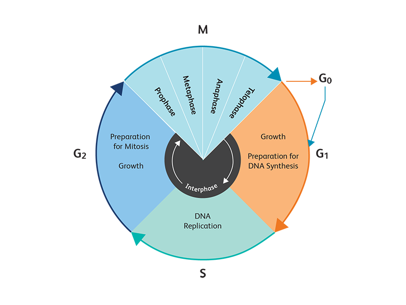
The cell cycle has two major phases: interphase, the phase between mitotic events, and the mitotic phase, where the mother cell divides into two genetically identical daughter cells. Interphase has three distinct, successive stages. During the first stage called G1, cells monitor their environment and, when the requisite signals are received, the cells synthesize RNA and proteins to induce growth. When conditions are right, cells enter the S stage of the cell cycle and commit to DNA synthesis and replicate their chromosomal DNA. Finally, in the G2 phase, cells continue to grow and prepare for mitosis.
BD Biosciences offers a wide variety of reagents and resources for cell cycle research.

Cell Proliferation
Cell proliferation is an increase in the number of cells as a result of growth and division. The balance of cell proliferation and apoptosis is important for both development and normal tissue homeostasis. A number of techniques are used to assess cell proliferation. During the synthesis (S) phase of the cell cycle, DNA polymerases incorporate a variety of nucleosides (deoxyadenosine, deoxyguanosine, deoxycytidine and thymidine) into the newly extending strands of DNA. Using analogs to these nucleosides provides a way to measure cell proliferation. 5-bromo-2’-deoxyuridine (BrdU), a thymidine analog, is widely used to measure de novo DNA synthesis. Several cell cycle-associated proteins, such as Ki-67, are also used as indicators of cell proliferation. Fluorescent or nonfluorescent cytoplasmic proliferation dyes can also be used as a measure cell proliferation.3

BD Biosciences offers BD Horizon™ Violet Proliferation Dye 450 and BD Horizon™ CFSE for the detection of cell proliferation with the violet laser and blue laser, respectively, which facilitates the use of larger panels. This allows the determination of more data from limited samples using multicolor flow cytometry.
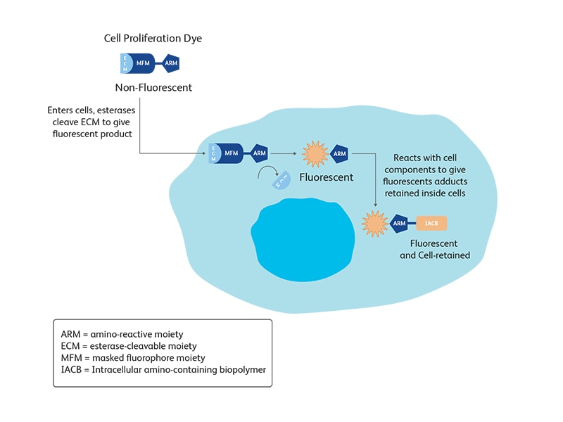
Both proliferation dyes are nonfluorescent esterified dyes. The ester group allows the dye to enter the cell. Once the dye is inside the cell, esterases cleave off the ester group to convert the dye into a fluorescent product and trap it inside the cell. With each replication event the amount of dye in the cell is decreased, leading to a characteristic pattern.
Apoptosis and Cell Death
Apoptosis is an organized process that signals cells to self-destruct for cell renewal or to control aberrant cell growth. As cells become damaged or are no longer needed, they undergo apoptosis or programmed cell death, a normal physiological process that occurs during embryonic development and tissue homeostasis. Apoptosis controls the orderly death of damaged cells, whereas necrosis occurs as a result of tissue damage, causing the loss of both damaged and surrounding cells.
The apoptotic process is characterized by certain morphological features. These include changes in the plasma membrane (such as loss of membrane symmetry and loss of membrane attachment), a condensation of the cytoplasm and nucleus, protein cleavage, and internucleosomal cleavage of DNA. In the final stages of the process, dying cells become fragmented into apoptotic bodies and consequently are eliminated by phagocytic cells without significant inflammatory damage to surrounding cells.

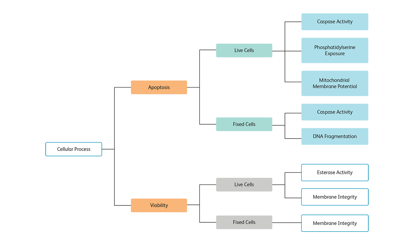
Methods for detecting apoptosis or dead cells (viability) by cell preparation type.
However, some cell types do not display characteristic features of apoptosis. In those cases, multiple aspects of apoptosis might need to be analyzed to confirm the mechanism of cell death.
To support this spectrum of requirements, BD Biosciences offers a full range of apoptosis detection tools and technologies for measuring indicators at different stages across the apoptotic process.
References
- Pack LR, Daigh LH, Meyer T. Putting the brakes on the cell cycle: mechanisms of cellular growth arrest. Curr Opin Cell Biol. 2019; 60:106-113. doi: 10.1016/j.ceb.2019.05.005
- Pistritto G, Triscuiuoglio D, Ceci C, et al. Apoptosis as anticancer mechanism: function and dysfunction of its modulators and targeted therapeutic strategies. Aging. 2016;8(4):603-619.
doi: 10.18632/aging.100934
- Romar G, Kupper T, Divito S. Research techniques made simple: Techniques to assess cell proliferation. J of Investigative Dermatol. 2016;136(7):E1-E7.
For Research Use Only. Not for use in diagnostic or therapeutic procedures.