
Preparation And Storage
Recommended Assay Procedures
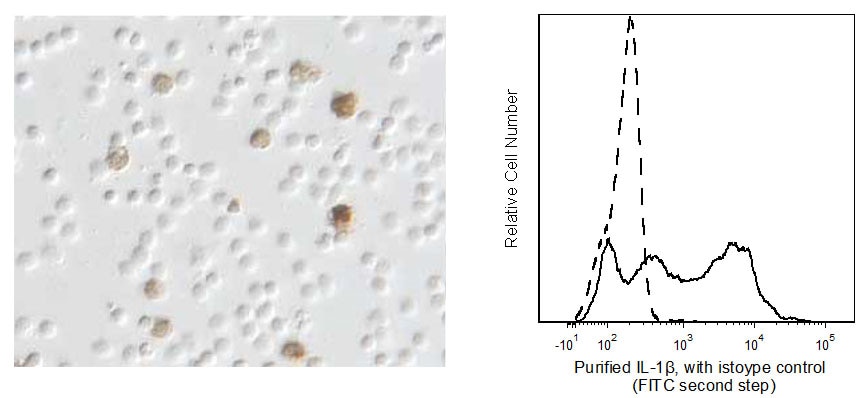
Immunocytochemistry: The purified format of the AS10 (Cat. No. 550007) antibody can be used to identify and enumerate human IL-1β producing cells by immunocytochemistry. For optimal indirect immunocytochemical staining, the AS10 antibody should be titrated (≤ 1 µg) and visualized via a three-step staining procedure in combination with Biotin Goat Anti-Mouse IgG, Streptavidin-HRP, and DAB substrate. Please see protocol for a detailed description of the immunocytochemical procedure. For optimal detection of cytokine producing cells, Pharmingen recommends horseradish peroxidase as the preferred enzyme system.
CYTOKINE IMMUNOCYTOCHEMISTRY PROTOCOL
REAGENTS REQUIRED
1. Fixation Buffer: 5% formalin (10% formalin, CMS, Cat. No. 245-684) is dissolved in phosphate buffered-saline (PBS) (Bacto FA Buffer, Difco Laboratories, Cat. No. 2314-15-0), or BD Pharmingen™ ICC Fixation Buffer (BD Cat. No. 550010)
2. Endogenous Peroxidase Blocking Buffer: DAKO Peroxidase Blocking Reagent (DAKO, Cat. No. S2001).
3. Endogenous Biotin Blocking Buffer: Biotin/Avidin Blocking Kit (Vector Laboratories, Cat. No. SP-2001).
4. Antibody dilution buffer: BD™ Pharmingen Antibody Diluent for IHC (Cat. No. 559148) supplemented with saponin.
5. Microscopic slides: Adhesion Slides (Erie Scientific Company, Cat. No. ER-202B-AD) or for cytospins, Colorfrost /Plus slides (Fisher, Cat. No. 12-550-17).
6. Biotin Goat anti-Mouse IgG (Cat. No. 550337) or the Anti-Mouse Ig HRP Detection Kit (Cat. No. 551011).
7. Detection system: BD Pharmingen Streptavidin-horseradish peroxidase (HRP), (Cat. No. 550946) or the Anti-Mouse Ig HRP Detection Kit (Cat. No. 551011).
8. Mounting medium for short-term storage: Aqua-mount® (Lerner Laboratories, Cat. No. 13800).
9. DAB Substrate Kit (contains 3-3 -Diaminobenzidine tetra hydrochloride), (BD Cat. No. 550880) or Anti-Mouse Ig HRP Detection Kit (Cat. No. 551011).
PROCEDURE FOR IMMUNOCYTOCHEMICAL STAINING OF SINGLE-CELL PREPARATIONS
This procedure describes the immunoenzymatic technique of staining cytokines within individual cells that are immobilized on microscopic slides via adherence (adherent slides) or centrifugation (cytospins).
ADHESION SLIDES
1. Harvest cells and wash them twice in PBS using centrifugation (400 x g for 5 min) to remove residual protein.
2. Adjust the cell concentration at 4-5 x 10e6 cells/ml in PBS.
3. Place 20 µl of the cell suspension in each well of the adhesion slides and let them adhere at room temperature (RT) for 20 min. Please note that the slides should be washed in PBS at RT for 5 min before transferring the cells.
4. Fix cells on slides using fixation buffer for 15 min at RT.
5. Wash slides 2X in PBS with 5 min incubations.
6. Block slides with PBS supplemented with 1% (w/v) BSA (Sigma) for 30 min at RT or 10 min at 37°C.
7. Wash slides 2X in PBS and proceed with staining or air dry them and store them at -80°C for future use.
8. Incubate slides with 20 µl of 1% goat serum and PBS with 0.1% (w/v) saponin for 30 min at RT.
9. Wash slides 2X with PBS with 5 min incubations.
10. Block endogenous peroxidase activity with Endogenous Peroxidase Blocking Buffer (20 µl/well) for 10 min at RT.
11. Wash 2X in PBS with 5 min incubations.
12. Incubate each well with Avidin (20 µl/well) for 15 min.
13. Wash 2X in PBS with 5 min incubations.
14. Incubate each well with Biotin (20 µl/well) for 15 min.
15. Wash 2X in PBS with 5 min incubations.
16. Incubate each well for 1 hr at RT with 20 µl of purified cytokine-specific antibody or appropriate immunoglobulin isotype control diluted in Pharmingen's IHC Diluent Buffer supplemented with saponin.
17. Wash slides 2X in PBS with 5 min incubations.
18. Incubate each well with 20 µl of a biotinylated secondary antibody diluted in IHC Cytokine Diluent Buffer for 30 min at RT.
19. Wash 2X in PBS with 5 min incubations.
20. Apply 20 µl of Streptavidin-HRP (BD Cat. No. 550946) to each well on slides and incubate for 30 min at RT.
21. Wash slides 2X with PBS with 5 minutes incubations.
22. Incubate with DAB Substrate as directed, (BD Cat. No. 550880) for less than 5 min at RT.
23. Stop the development of the color reaction by washing with PBS.
24. The slides are subsequently mounted in short-term storage mounding medium.
CYTOSPINS
1. Assemble the Cytospin's sample chamber (e.g. Cytospin 3, Shandon, UK or comparable centrifuge), filter card, slide and cytospin racks according to manufacturer's specifications.
2. Load 40 µl of approximately 1 x 10e6 cells to each sample chamber.
3. Spin slides at 600 rpm for 2 min.
4. Take slides out of the cytospin rack and place them on a staining rack.
5. For fixation and staining please follow the steps 4 through 24 specified above for staining cells on adhesion slides.
Product Notices
- Since applications vary, each investigator should titrate the reagent to obtain optimal results.
- An isotype control should be used at the same concentration as the antibody of interest.
- Caution: Sodium azide yields highly toxic hydrazoic acid under acidic conditions. Dilute azide compounds in running water before discarding to avoid accumulation of potentially explosive deposits in plumbing.
- Sodium azide is a reversible inhibitor of oxidative metabolism; therefore, antibody preparations containing this preservative agent must not be used in cell cultures nor injected into animals. Sodium azide may be removed by washing stained cells or plate-bound antibody or dialyzing soluble antibody in sodium azide-free buffer. Since endotoxin may also affect the results of functional studies, we recommend the NA/LE (No Azide/Low Endotoxin) antibody format, if available, for in vitro and in vivo use.
- Please refer to www.bdbiosciences.com/us/s/resources for technical protocols.
Companion Products


The AS10 antibody reacts with human interleukin-1β (IL-1β) which is also known as endogenous pyrogen (EP), leukocyte endogenous mediator (LEM), mononuclear cell factor (MCF) and lymphocyte-activating factor (LAF). IL-1β is a proinflammatory cytokine that is synthesized as a precursor of 31 kDa and is converted intracellularly to the mature 17.5 kDa form, after cleavage by the IL-1β-converting enzyme (ICE). In healthy individuals, IL-1β is secreted non-constitutively by blood monocytes, tissue macrophages and dendritic cells. IL-1β is also constitutively expressed in the human hypothalamus. Many malignant tumors express IL-1β as part of their neoplastic nature. The AS10 antibody has been reported not to recognize human IL-1α nor cross react with mouse IL-1β.
Development References (2)
-
Hsu SM, Raine L, Fanger H. A comparative study of the peroxidase-antiperoxidase method and an avidin-biotin complex method for studying polypeptide hormones with radioimmunoassay antibodies. Am J Clin Pathol. 1981; 75(5):734-738. (Methodology). View Reference
-
Hsu SM, Raine L, Fanger H. Use of avidin-biotin-peroxidase complex (ABC) in immunoperoxidase techniques: a comparison between ABC and unlabeled antibody (PAP) procedures. J Histochem Cytochem. 1981; 29(4):577-580. (Methodology). View Reference
Please refer to Support Documents for Quality Certificates
Global - Refer to manufacturer's instructions for use and related User Manuals and Technical data sheets before using this products as described
Comparisons, where applicable, are made against older BD Technology, manual methods or are general performance claims. Comparisons are not made against non-BD technologies, unless otherwise noted.
For Research Use Only. Not for use in diagnostic or therapeutic procedures.
Refer to manufacturer's instructions for use and related User Manuals and Technical Data Sheets before using this product as described.
Comparisons, where applicable, are made against older BD technology, manual methods or are general performance claims. Comparisons are not made against non-BD technologies, unless otherwise noted.