
Preparation And Storage
Recommended Assay Procedures
BD™ CompBeads can be used as surrogates to assess fluorescence spillover (Compensation). When fluorochrome conjugated antibodies are bound to CompBeads, they have spectral properties very similar to cells. However, for some fluorochromes there can be small differences in spectral emissions compared to cells, resulting in spillover values that differ when compared to biological controls. It is strongly recommended that when using a reagent for the first time, users compare the spillover on cell and CompBead to ensure that BD Comp beads are appropriate for your specific cellular application.
For optimal and reproducible results, BD Horizon Brilliant Stain Buffer should be used anytime two or more BD Horizon Brilliant dyes are used in the same experiment. Fluorescent dye interactions may cause staining artifacts which may affect data interpretation. The BD Horizon Brilliant Stain Buffer was designed to minimize these interactions. More information can be found in the Technical Data Sheet of the BD Horizon Brilliant Stain Buffer (Cat. No. 563794/566349) or the BD Horizon Brilliant Stain Buffer Plus (Cat. No. 566385).
Product Notices
- Since applications vary, each investigator should titrate the reagent to obtain optimal results.
- An isotype control should be used at the same concentration as the antibody of interest.
- Caution: Sodium azide yields highly toxic hydrazoic acid under acidic conditions. Dilute azide compounds in running water before discarding to avoid accumulation of potentially explosive deposits in plumbing.
- For fluorochrome spectra and suitable instrument settings, please refer to our Multicolor Flow Cytometry web page at www.bdbiosciences.com/colors.
- Please refer to http://regdocs.bd.com to access safety data sheets (SDS).
- BD Horizon Brilliant Stain Buffer is covered by one or more of the following US patents: 8,110,673; 8,158,444; 8,575,303; 8,354,239.
- Please refer to www.bdbiosciences.com/us/s/resources for technical protocols.
Companion Products


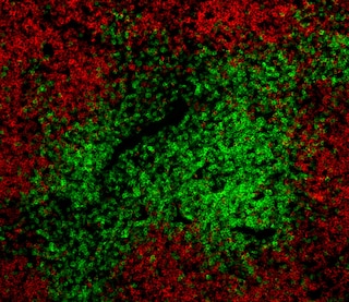
The G9P3 monoclonal antibody specifically recognizes mouse EVA1, which is encoded by the Mpzl2 gene. EVA1 is a member of the immunoglobulin-like cell-adhesion molecule (Ig-CAM or IgSF CAM) family, members of which are anchored to the surface of epithelial and endothelial cells via a GPI domain. This class of adhesion molecules interacts intracellularly with cytoskeletal proteins and extracellularly with Ig-CAM family members, by homophilic or heterophilic binding, and with other classes of adhesion molecules such as integrins and cadherins. EVA1 is expressed on and is involved in the structural integrity of epithelia throughout the body, eg mammary glands, liver, inner ear, and the blood-brain barrier. In the thymus, EVA1 is expressed both on thymic epithelial cells (TECs) and on a subset of developing thymocytes, known as DN3. It is a low-affinity mediator of developmental signals between thymocytes and TEC that support the differentiation and survival of both developing thymocytes and the TECs. G9P3 mAb was produced in EVA1 knockout mice immunized with recombinant mouse EVA1 extracellular region, and it recognizes mouse and human EVA1 expressed on transfected cells.

Development References (10)
-
Chatterjee G, Carrithers LM, Carrithers MD. Epithelial V-like antigen regulates permeability of the blood-CSF barrier.. Biochem Biophys Res Commun. 2008; 372(3):412-7. (Biology). View Reference
-
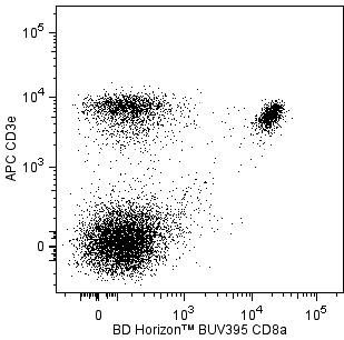
Garabatos N, Blanco J, Fandos C, et al. A monoclonal antibody against the extracellular domain of mouse and human epithelial V-like antigen 1 reveals a restricted expression pattern among CD4- CD8- thymocytes.. Monoclon Antib Immunodiagn Immunother. 2014; 33(5):305-11. (Immunogen: Flow cytometry). View Reference
-
Guttinger M, Sutti F, Panigada M, et al. Epithelial V-like antigen (EVA), a novel member of the immunoglobulin superfamily, expressed in embryonic epithelia with a potential role as homotypic adhesion molecule in thymus histogenesis.. J Cell Biol. 1998; 141(4):1061-71. (Biology). View Reference
-
Iacovelli S, Iosue I, Di Cesare S, Guttinger M. Lymphoid EVA1 expression is required for DN1-DN3 thymocytes transition. PLoS One. 2009; 4(10):e7586. (Biology). View Reference
-
Kurd N, Robey EA. T-cell selection in the thymus: a spatial and temporal perspective.. Immunol Rev. 2016; 271(1):114-26. (Biology). View Reference
-
Lin X, Cui M, Xu D, et al. Liver-specific deletion of Eva1a/Tmem166 aggravates acute liver injury by impairing autophagy. Cell Death Dis. 2018; 9(7):768. (Biology). View Reference
-
Litvinov IV, Netchiporouk E, Cordeiro B, et al.. Ectopic expression of embryonic stem cell and other developmental genes in cutaneous T-cell lymphoma. Oncoimmunology. 2014; 3(11):e970025. (Biology). View Reference
-
Ramena G, Yin Y, Yu Y, Walia V, Elble RC. CLCA2 Interactor EVA1 Is Required for Mammary Epithelial Cell Differentiation.. PLoS ONE. 2016; 11(3):e0147489. (Biology). View Reference
-
Wesdorp M, Murillo-Cuesta S, Peters T, et al. MPZL2, Encoding the Epithelial Junctional Protein Myelin Protein Zero-like 2, Is Essential for Hearing in Man and Mouse. Am J Hum Genet. 2018; 103(1):74-88. (Biology). View Reference
-
Wright E, Rahgozar K, Hallworth N, Lanker S, Carrithers MD. Epithelial V-like antigen mediates efficacy of anti-alpha₄ integrin treatment in a mouse model of multiple sclerosis.. PLoS ONE. 2013; 8(8):e70954. (Biology). View Reference
Please refer to Support Documents for Quality Certificates
Global - Refer to manufacturer's instructions for use and related User Manuals and Technical data sheets before using this products as described
Comparisons, where applicable, are made against older BD Technology, manual methods or are general performance claims. Comparisons are not made against non-BD technologies, unless otherwise noted.
For Research Use Only. Not for use in diagnostic or therapeutic procedures.
Refer to manufacturer's instructions for use and related User Manuals and Technical Data Sheets before using this product as described.
Comparisons, where applicable, are made against older BD technology, manual methods or are general performance claims. Comparisons are not made against non-BD technologies, unless otherwise noted.