Old Browser
Looks like you're visiting us from {countryName}.
Would you like to stay on the current country site or be switched to your country?
BD Pharmingen™ PE Mouse Anti-Mouse CD66a (CEACAM1a)
Clone CC1 (also known as MAb-CCl; mAb CC1)
(RUO)

.png)

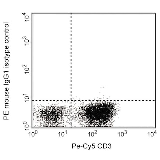
Multicolor flow cytometric analysis of CD66a (CEACAM1a) expression on viable mouse splenic leucocytes. BALB/c mouse spleen cells were treated with BD Pharm Lyse™ Lysing Buffer (Cat. No. 555899) to lyse erythrocytes, washed, and preincubated with Purified Rat Anti-Mouse CD16/CD32 antibody (Mouse BD Fc Block™) (Cat. No. 553141/553142). The cells were then stained with APC Rat Anti-Mouse CD45R/B220 antibody (Cat. No. 561880/553092) and either PE Mouse IgG1, κ Isotype Control (Cat. No. 554680; Left Plot) or PE Mouse Anti-CD66a (CEACAM1a) antibody (Cat. No. 566828; Right Plot) at 0.25 µg/test. DAPI (4',6-Diamidino-2-Phenylindole, Dihydrochloride) Solution (Cat. No. 564907) was added to cells right before analysis. The pseudocolor density plot showing the correlated expression of CD66a (CEACAM1) [or Ig Isotype control staining] versus CD45R/B220 was derived from gated events with the forward and side light-scatter characteristics of viable (DAPI-negative) cells. Flow cytometry and data analysis were performed using a BD LSRFortessa™ Cell Analyzer System and FlowJo™ software. Data shown on this Technical Data Sheet are not lot specific.
.png)

BD Pharmingen™ PE Mouse Anti-Mouse CD66a (CEACAM1a)
.png)
Regulatory Status Legend
Any use of products other than the permitted use without the express written authorization of Becton, Dickinson and Company is strictly prohibited.
Preparation And Storage
Product Notices
- Since applications vary, each investigator should titrate the reagent to obtain optimal results.
- An isotype control should be used at the same concentration as the antibody of interest.
- Caution: Sodium azide yields highly toxic hydrazoic acid under acidic conditions. Dilute azide compounds in running water before discarding to avoid accumulation of potentially explosive deposits in plumbing.
- For fluorochrome spectra and suitable instrument settings, please refer to our Multicolor Flow Cytometry web page at www.bdbiosciences.com/colors.
- Please refer to http://regdocs.bd.com to access safety data sheets (SDS).
- Please refer to www.bdbiosciences.com/us/s/resources for technical protocols.
Companion Products


The CC1 monoclonal antibody specifically recognizes carcinoembryonic antigen-related cell adhesion molecule 1a (CEACAM1a or CEACAM1[a]), an allotypic form of CEACAM1 which is also known as CD66a, Murine hepatitis virus receptor (MHV-R), or Biliary glycoprotein 1 (BGP-1). Four known isoforms of mouse CD66a arise from alternative splicing of RNA transcripts encoded by Ceacam1, a member of the carcinoembryonic antigen (CEA) family and Ig gene superfamily. These isoforms are type I transmembrane proteins that include a heavily glycosylated extracellular region with an N-terminal IgV-like domain and up to three IgC2-like domains followed by a transmembrane region and a cytoplasmic tail of relatively short (10 amino acids) or long (73 amino acids) length. The cytoplasmic tails enable interactions with other intracellular molecules to initiate or regulate cellular responses. The two CD66a isoforms with long cytoplasmic tails contain immunoreceptor tyrosine-based inhibitory motifs (ITIMs) that may enable CD66a to function as an immune checkpoint inhibitor. CD66a is expressed on a variety of cell types including certain epithelial cells, endothelial cells, B cells, activated T cells, NK cells, monocytes, dendritic cells (DC), and neutrophils. CD66a (CEACAM1a) is a multifunctional protein. Through their IgV-like domain, CD66a (CEACAM1a) molecules function as homophilic and heterophilic intercellular adhesion molecules. They can also function as MHV-Rs, angiogenic factors, regulators of cellular proliferation and differentiation, and tumor suppressors. Two distinct Ceacam1 alleles, (a and b), exist because of differences in their IgV-like domain gene sequences. Ceacam1a is found in most inbred mouse strains including BALB/c, C57BL/6, and C3H mice whereas Ceacam1b is found in SJL mice. CD66a (CEACAM1a) proteins are specifically bound by the CC1 antibody whereas CEACAM1b proteins are not. The CC1 antibody recognizes an epitope in the N-terminal domain of mouse CD66a (CEACAM1a) proteins.

Development References (10)
-
Chen Z, Chen L, Baker K, et al. CEACAM1 dampens antitumor immunity by down-regulating NKG2D ligand expression on tumor cells. J Exp Med. 2011; 208(13):2633-2640. (Clone-specific: Flow cytometry). View Reference
-
Dveksler GS, Dieffenbach CW, Cardellichio CB, et al. J Virol. 1993; 67(1):1-8. (Clone-specific). View Reference
-
Dveksler GS, Pensiero MN, Cardellichio CB, et al. Cloning of the mouse hepatitis virus (MHV) receptor: expression in human and hamster cell lines confers susceptibility to MHV. J Virol. 1991; 65(12):6881-6891. (Clone-specific: Blocking). View Reference
-
Gray-Owen SD, Blumberg RS. CEACAM1: contact-dependent control of immunity.. Nat Rev Immunol. 2006; 6(6):433-46. (Biology). View Reference
-
Hemmila E, Turbide C, Olson M, Jothy S, Holmes KV, Beauchemin N. Ceacam1a-/- mice are completely resistant to infection by murine coronavirus mouse hepatitis virus A59. J Virol. 2004; 78(18):10156-10165. (Clone-specific). View Reference
-
Holmes KV, Dveksler G, Gagneten S, et al. Coronavirus receptor specificity.. Adv Exp Med Biol. 1993; 342:261-6. (Clone-specific: Immunoaffinity chromatography). View Reference
-
Khairnar V, Duhan V, Maney SK, et al. CEACAM1 induces B-cell survival and is essential for protective antiviral antibody production.. Nat Commun. 2015; 6:6217. (Clone-specific: Flow cytometry). View Reference
-
Nakajima A, Iijima H, Neurath MF, et al. Activation-induced expression of carcinoembryonic antigen-cell adhesion molecule 1 regulates mouse T lymphocyte function.. J Immunol. 2002; 168(3):1028-35. (Clone-specific: Flow cytometry). View Reference
-
Williams RK, Jiang GS, Holmes KV.. Receptor for mouse hepatitis virus is a member of the carcinoembryonic antigen family of glycoproteins. Proc Natl Acad Sci U S A. 1991; 88(13):5533-5536. (Clone-specific: Immunoaffinity chromatography, Radioimmunoassay). View Reference
-
Williams RK, Jiang GS, Snyder SW, Frana MF, Holmes KV.. Purification of the 110-kilodalton glycoprotein receptor for mouse hepatitis virus (MHV)-A59 from mouse liver and identification of a nonfunctional, homologous protein in MHV-resistant SJL/J mice. J Virol. 1990; 64(8):3817-3823. (Immunogen: Blocking, ELISA, Immunoaffinity chromatography, Inhibition, Radioimmunoassay). View Reference
Please refer to Support Documents for Quality Certificates
Global - Refer to manufacturer's instructions for use and related User Manuals and Technical data sheets before using this products as described
Comparisons, where applicable, are made against older BD Technology, manual methods or are general performance claims. Comparisons are not made against non-BD technologies, unless otherwise noted.
For Research Use Only. Not for use in diagnostic or therapeutic procedures.
Refer to manufacturer's instructions for use and related User Manuals and Technical Data Sheets before using this product as described.
Comparisons, where applicable, are made against older BD technology, manual methods or are general performance claims. Comparisons are not made against non-BD technologies, unless otherwise noted.