Dear Customer, If you experience any text readability or display issues on our website, please update your web browser to the latest version
.
See More
Dear Customer, If you experience any text readability or display issues on our website, please update your web browser to the latest version
.
See MoreDear Customer, If you experience any text readability or display issues on our website, please update your web browser to the latest version
.
Looks like you're visiting us from {countryName}.
Would you like to stay on the current location site or be switched to your location?
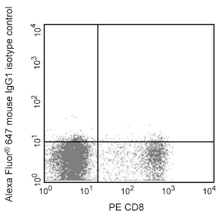
Two-color flow cytometric analysis of MCP-1 expression in human periphereal blood mononuclear cells. Human PBMC were cultured (6 hr) with Recombinant Human IFN-γ (Cat. No. 554617/554616; 10 ng/ml) and lipopolysaccharide (Sigma, Cat. No. L-8274; 1.0 μg/ml) in the presence of BD GolgiStop™ Protein Transport Inhibitor (containing Monensin) (Cat. No. 554724). The cells were harvested, washed with BD Pharmingen™ Stain Buffer (FBS) (Cat. No. 554656) and stained with FITC Mouse Anti-Human CD14 antibody (Cat. No. 555397/557153/561712). After washing, the cells were fixed and permeabilized with BD Cytofix/Cytoperm™ Fixation and Permeabilization Solution (Cat. No. 554722). The cells were then stained in BD Perm/Wash™ Buffer (Cat. No. 554723) with either Alexa Fluor® 647 Mouse IgG1 κ Isotype Control (Cat. No. 557732; Left Panel) or Alexa Fluor® 647 Mouse Anti-Human MCP-1 antibody (Cat. No. 563496; Right Panel) using BD Biosciences Intracellular Cytokine Staining protocol. Two-color flow cytometric dot plots show the correlated expression patterns of MCP-1 (or Ig Isotype control staining) versus CD14 for gated events with the forward and side light-scatter characteristics of intact stimulated PBMC. Flow cytometric analysis was performed using a BD™ LSR II Flow Cytometer System.
Any use of products other than the permitted use without the express written authorization of Becton, Dickinson and Company is strictly prohibited.
The 5D3-F7 monoclonal antibody specifically binds to human monocyte chemoattractant protein-1 (MCP-1), also known as CCL2 (C-C motif chemokine 2), Monocyte chemotactic and activating factor (MCAF), and Small-inducible cytokine A2 (SCYA2). MCP-1 is a 10-14 kDa glycoprotein member of the beta or CC family of chemokines. It expressed by monocytes, fibroblasts, endothelial cells and other cell types in response to IL-1, IL-6, TNF, and a variety of other stimuli. MCP-1 binds to and exerts its biological activity through G-protein coupled chemokine receptors including CCR2/CD192 and CCR4/CD194. It serves as a chemoattractant and activating factor for monocytes and other cell types including T cells, NK cells, and basophils.
MCP-1 is a member of the CC chemokine family and it is produced by monocytes, T lymphocytes, fibroblasts, endothelial cells, smooth muscle cells, keratinocytes and some tumors. Its production can be induced by LPS or IFN-γ. Clone 5D3-F7 also cross reacts with an intracellular component of LPS-stimulated (24 hours) peripheral blood monocytes of rhesus and cynomolgus macaque monkeys. The staining pattern observed on non-human primate monocytes is not as strong as that seen on normal human peripheral blood monocytes.
Please refer to Support Documents for Quality Certificates
Global - Refer to manufacturer's instructions for use and related User Manuals and Technical data sheets before using this products as described
Comparisons, where applicable, are made against older BD Technology, manual methods or are general performance claims. Comparisons are not made against non-BD technologies, unless otherwise noted.
For Research Use Only. Not for use in diagnostic or therapeutic procedures.
Refer to manufacturer's instructions for use and related User Manuals and Technical Data Sheets before using this product as described.
Comparisons, where applicable, are made against older BD technology, manual methods or are general performance claims. Comparisons are not made against non-BD technologies, unless otherwise noted.