







Preparation And Storage


Recommended Assay Procedures
For optimal and reproducible results, BD Horizon Brilliant Stain Buffer should be used anytime two or more BD Horizon Brilliant dyes are used in the same experiment. Fluorescent dye interactions may cause staining artifacts which may affect data interpretation. The BD Horizon Brilliant Stain Buffer was designed to minimize these interactions. More information can be found in the Technical Data Sheet of the Brilliant Stain Buffer (Cat. No. 563794/566349) or the Brilliant Stain Buffer Plus (Cat. No. 566385).
Product Notices
- Since applications vary, each investigator should titrate the reagent to obtain optimal results.
- An isotype control should be used at the same concentration as the antibody of interest.
- Source of all serum proteins is from USDA inspected abattoirs located in the United States.
- Caution: Sodium azide yields highly toxic hydrazoic acid under acidic conditions. Dilute azide compounds in running water before discarding to avoid accumulation of potentially explosive deposits in plumbing.
- For fluorochrome spectra and suitable instrument settings, please refer to our Multicolor Flow Cytometry web page at www.bdbiosciences.com/colors.
- Brilliant Violet™ 510 is a trademark of Sirigen.
- Although hamster immunoglobulin isotypes have not been well defined, BD Biosciences Pharmingen has grouped Armenian and Syrian hamster IgG monoclonal antibodies according to their reactivity with a panel of mouse anti-hamster IgG mAbs. A table of the hamster IgG groups, Reactivity of Mouse Anti-Hamster Ig mAbs, may be viewed at http://www.bdbiosciences.com/documents/hamster_chart_11x17.pdf.
- BD Horizon Brilliant Stain Buffer is covered by one or more of the following US patents: 8,110,673; 8,158,444; 8,575,303; 8,354,239.
- BD Horizon Brilliant Violet 510 is covered by one or more of the following US patents: 8,575,303; 8,354,239.
- Please refer to www.bdbiosciences.com/us/s/resources for technical protocols.
Companion Products
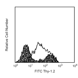



The GL3 monoclonal antibody specifically binds to a common epitope of the δ chain of the T-cell Receptor (TCR) complex on γδ TCR-expressing T lymphocytes and NK-T cells of all mouse strains tested. It does not react with αβ TCR-bearing T cells. In the mouse, cells expressing the γδ TCR are found in the thymus, intestinal epithelium, epidermis, dermis, pulmonsry epithelium, peritoneum, liver, and peripheral lymphoid organs.
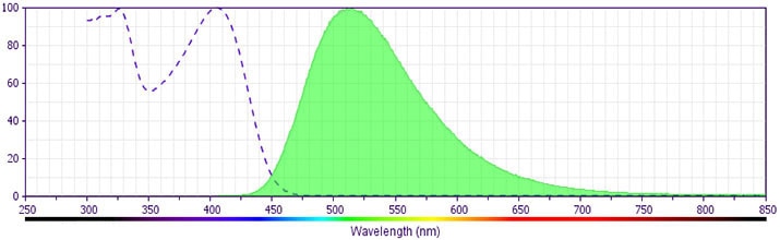
The antibody was conjugated to BD Horizon™ BV510 which is part of the BD Horizon™ Brilliant Violet™ family of dyes. With an Ex Max of 405-nm and Em Max at 510-nm, BD Horizon™ BV510 can be excited by the violet laser and detected in the BD Horizon™ V500 (525/50-nm) filter set. BD Horizon™ BV510 conjugates are useful for the detection of dim markers off the violet laser.
Development References (15)
-
Goodman T, LeCorre R, Lefrancois L. A T-cell receptor gamma delta-specific monoclonal antibody detects a V gamma 5 region polymorphism. Immunogenetics. 1992; 35(1):65-68. (Clone-specific: Flow cytometry). View Reference
-
Goodman T, Lefrancois L. Intraepithelial lymphocytes. Anatomical site, not T cell receptor form, dictates phenotype and function. J Exp Med. 1989; 170(5):1569-1581. (Clone-specific: Flow cytometry, Immunoprecipitation). View Reference
-
Kaufmann SH, Blum C, Yamamoto S. Crosstalk between alpha/beta T cells and gamma/delta T cells in vivo: activation of alpha/beta T-cell responses after gamma/delta T-cell modulation with the monoclonal antibody GL3. Proc Natl Acad Sci U S A. 1993; 90(20):9620-9624. (Clone-specific: Depletion). View Reference
-
King DP, Hyde DM, Jackson KA, et al. Cutting edge: protective response to pulmonary injury requires gamma delta T lymphocytes. J Immunol. 1999; 162(9):5033-5036. (Clone-specific: Flow cytometry). View Reference
-
Lefrancois L, Barrett TA, Havran WL, Puddington L. Developmental expression of the alpha IEL beta 7 integrin on T cell receptor gamma delta and T cell receptor alpha beta T cells. Eur J Immunol. 1994; 24(3):635-640. (Clone-specific: Immunohistochemistry). View Reference
-
Lefrancois L. Phenotypic complexity of intraepithelial lymphocytes of the small intestine. J Immunol. 1991; 147(6):1746-1751. (Clone-specific). View Reference
-
MacDonald HR, Schreyer M, Howe RC, Bron C. Selective expression of CD8 alpha (Ly-2) subunit on activated thymic gamma/delta cells. Eur J Immunol. 1990; 20(4):927-930. (Biology: Flow cytometry). View Reference
-
Nakazawa S, Brown AE, Maeno Y, Smith CD, Aikawa M. Malaria-induced increase of splenic gamma delta T cells in humans, monkeys, and mice. 1994; 79(3):391-398. (Clone-specific: Immunohistochemistry). View Reference
-
Shinohara K, Ikarashi Y, Maruoka H, et al. Functional and phenotypical characteristics of hepatic NK-like T cells in NK1.1-positive and -negative mouse strains. Eur J Immunol. 1999; 29(6):1871-1878. (Clone-specific: Flow cytometry). View Reference
-
Skeen MJ, Ziegler HK. Induction of murine peritoneal gamma/delta T cells and their role in resistance to bacterial infection. J Exp Med. 1993; 178(3):971-984. (Clone-specific: Flow cytometry, In vivo exacerbation). View Reference
-
Tamaki K, Yasaka N, Chang CH, et al. Identification and characterization of novel dermal Thy-1 antigen-bearing dendritic cells in murine skin. J Invest Dermatol. 1996; 106(3):571-575. (Clone-specific: Fluorescence microscopy, Immunofluorescence, Immunohistochemistry). View Reference
-
Tigelaar RE, Lewis JM, Bergstresser PR. TCR gamma/delta+ dendritic epidermal T cells as constituents of skin-associated lymphoid tissue. J Invest Dermatol. 1990; 94(6):58S-63S. (Biology). View Reference
-
Vicari AP, Mocci S, Openshaw P, O'Garra A, Zlotnik A. Mouse gamma delta TCR+NK1.1+ thymocytes specifically produce interleukin-4, are major histocompatibility complex class I independent, and are developmentally related to alpha beta TCR+NK1.1+ thymocytes. Eur J Immunol. 1996; 26(7):1424-1429. (Clone-specific: Flow cytometry, Fluorescence activated cell sorting). View Reference
-
Yanez DM, Batchelder J, van der Heyde HC, Manning DD, Weidanz WP. Gamma delta T-cell function in pathogenesis of cerebral malaria in mice infected with Plasmodium berghei ANKA. Infect Immun. 1999; 67(1):446-448. (Clone-specific: Depletion). View Reference
-
van der Heyde HC, Elloso MM, Chang WL, Kaplan M, Manning DD, Weidanz WP. Gamma delta T cells function in cell-mediated immunity to acute blood-stage Plasmodium chabaudi adami malaria. J Immunol. 1995; 154(8):3985-3990. (Clone-specific: Depletion). View Reference
Please refer to Support Documents for Quality Certificates
Global - Refer to manufacturer's instructions for use and related User Manuals and Technical data sheets before using this products as described
Comparisons, where applicable, are made against older BD Technology, manual methods or are general performance claims. Comparisons are not made against non-BD technologies, unless otherwise noted.
For Research Use Only. Not for use in diagnostic or therapeutic procedures.
