-
Reagents
- Flow Cytometry Reagents
-
Western Blotting and Molecular Reagents
- Immunoassay Reagents
-
Single-Cell Multiomics Reagents
- BD® OMICS-Guard Sample Preservation Buffer
- BD® AbSeq Assay
- BD® OMICS-One Immune Profiler Protein Panel
- BD® Single-Cell Multiplexing Kit
- BD Rhapsody™ ATAC-Seq Assays
- BD Rhapsody™ Whole Transcriptome Analysis (WTA) Amplification Kit
- BD Rhapsody™ TCR/BCR Next Multiomic Assays
- BD Rhapsody™ Targeted mRNA Kits
- BD Rhapsody™ Accessory Kits
- BD® OMICS-One Protein Panels
-
Functional Assays
-
Microscopy and Imaging Reagents
-
Cell Preparation and Separation Reagents
-
Thought Leadership
- Product News
- Blogs
-
Scientific Publications
-
Events
- Expanding PARADIGM to Infectious Disease Modeling: HIV & Tuberculosis
- CYTO 2023: Advancing the World of Cytometry
- Advances in Immune Monitoring Series
- Validating Flow Cytometry Assays for Cell Therapy
- Enhancing Cell Analysis with a New Set of Eyes
- BD Biosciences at International Clinical Cytometry Society 2025
Old Browser
This page has been recently translated and is available in French now.
Looks like you're visiting us from {countryName}.
Would you like to stay on the current country site or be switched to your country?
BD Pharmingen™ Purified Mouse Anti-Human CEACAM5 (CD66e)
Clone CB30 (RUO)

Immunohistochemical analysis of CEACAM5 (CD66e) expression
Left Panel - Immunohistochemical staining of Human Normal Colon. Formalin-fixed, paraffin-embedded sections of Human Normal Colon were stained with either Purified Mouse IgG1, κ Isotype Control (Cat. No. 554121; Left Image) or Purified Mouse Anti-CEACAM5 (CD66e) antibody (Cat. No. 568709; Right Image) followed by Biotin Goat Anti-Mouse Ig (Multiple Adsorption) [Cat. No. 550337] and Streptavidin HRP (Cat. No.550946). Antigen retrieval with BD Retrievagen A (Cat. No. 550524) was performed prior to staining. Original magnification 20x.
Right Panel - Immunohistochemical staining of Human Colon Cancer. Formalin-fixed, paraffin-embedded sections of Human Colon Cancer were stained with either Purified Mouse IgG1, κ Isotype Control (Cat. No. 554121; Left Image) or Purified Mouse Anti-CEACAM5 (CD66e) antibody (Cat. No. 568709; Right Image) followed by Biotin Goat Anti-Mouse Ig (Multiple Adsorption) [Cat. No. 550337] and Streptavidin HRP (Cat. No.550946). Antigen retrieval with BD Retrievagen A (Cat. No. 550524) was performed prior to staining. Original magnification 20x.

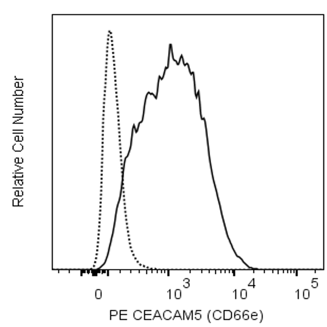
Flow cytometric analysis of CEACAM5 (CD66e) expression on BxPC-3 cells. Cells from the human BxPC-3 (Adenocarcinoma, ATCC CRL-1687™) cell line were stained with either Purified Mouse IgG1, κ Isotype Control (Cat. No. 554121; dotted line histogram) or Purified Mouse Anti-CEACAM5 (CD66e) antibody (Cat. No. 568709; solid line histogram) at 0.5 µg/test, followed by Biotin Goat Anti-Mouse Ig (Cat. No. 550337/553999) at 0.25 µg/test followed by PE Streptavidin (Cat. No. 554061). DAPI (4',6-Diamidino-2-Phenylindole, Dihydrochloride) Solution (Cat. No. 564907) was added to cells right before analysis. The fluorescence histogram showing CEACAM5 (CD66e) expression (or Ig Isotype control staining) was derived from gated events with the light-scatter characteristics of viable (DAPI-negative) cells. Flow cytometry and data analysis were performed using a BD LSRFortessa™ X20 Cell Analyzer System and FlowJo™ software.


Immunohistochemical analysis of CEACAM5 (CD66e) expression
Left Panel - Immunohistochemical staining of Human Normal Colon. Formalin-fixed, paraffin-embedded sections of Human Normal Colon were stained with either Purified Mouse IgG1, κ Isotype Control (Cat. No. 554121; Left Image) or Purified Mouse Anti-CEACAM5 (CD66e) antibody (Cat. No. 568709; Right Image) followed by Biotin Goat Anti-Mouse Ig (Multiple Adsorption) [Cat. No. 550337] and Streptavidin HRP (Cat. No.550946). Antigen retrieval with BD Retrievagen A (Cat. No. 550524) was performed prior to staining. Original magnification 20x.
Right Panel - Immunohistochemical staining of Human Colon Cancer. Formalin-fixed, paraffin-embedded sections of Human Colon Cancer were stained with either Purified Mouse IgG1, κ Isotype Control (Cat. No. 554121; Left Image) or Purified Mouse Anti-CEACAM5 (CD66e) antibody (Cat. No. 568709; Right Image) followed by Biotin Goat Anti-Mouse Ig (Multiple Adsorption) [Cat. No. 550337] and Streptavidin HRP (Cat. No.550946). Antigen retrieval with BD Retrievagen A (Cat. No. 550524) was performed prior to staining. Original magnification 20x.
Flow cytometric analysis of CEACAM5 (CD66e) expression on BxPC-3 cells. Cells from the human BxPC-3 (Adenocarcinoma, ATCC CRL-1687™) cell line were stained with either Purified Mouse IgG1, κ Isotype Control (Cat. No. 554121; dotted line histogram) or Purified Mouse Anti-CEACAM5 (CD66e) antibody (Cat. No. 568709; solid line histogram) at 0.5 µg/test, followed by Biotin Goat Anti-Mouse Ig (Cat. No. 550337/553999) at 0.25 µg/test followed by PE Streptavidin (Cat. No. 554061). DAPI (4',6-Diamidino-2-Phenylindole, Dihydrochloride) Solution (Cat. No. 564907) was added to cells right before analysis. The fluorescence histogram showing CEACAM5 (CD66e) expression (or Ig Isotype control staining) was derived from gated events with the light-scatter characteristics of viable (DAPI-negative) cells. Flow cytometry and data analysis were performed using a BD LSRFortessa™ X20 Cell Analyzer System and FlowJo™ software.

Immunohistochemical analysis of CEACAM5 (CD66e) expression
Left Panel - Immunohistochemical staining of Human Normal Colon. Formalin-fixed, paraffin-embedded sections of Human Normal Colon were stained with either Purified Mouse IgG1, κ Isotype Control (Cat. No. 554121; Left Image) or Purified Mouse Anti-CEACAM5 (CD66e) antibody (Cat. No. 568709; Right Image) followed by Biotin Goat Anti-Mouse Ig (Multiple Adsorption) [Cat. No. 550337] and Streptavidin HRP (Cat. No.550946). Antigen retrieval with BD Retrievagen A (Cat. No. 550524) was performed prior to staining. Original magnification 20x.
Right Panel - Immunohistochemical staining of Human Colon Cancer. Formalin-fixed, paraffin-embedded sections of Human Colon Cancer were stained with either Purified Mouse IgG1, κ Isotype Control (Cat. No. 554121; Left Image) or Purified Mouse Anti-CEACAM5 (CD66e) antibody (Cat. No. 568709; Right Image) followed by Biotin Goat Anti-Mouse Ig (Multiple Adsorption) [Cat. No. 550337] and Streptavidin HRP (Cat. No.550946). Antigen retrieval with BD Retrievagen A (Cat. No. 550524) was performed prior to staining. Original magnification 20x.

Flow cytometric analysis of CEACAM5 (CD66e) expression on BxPC-3 cells. Cells from the human BxPC-3 (Adenocarcinoma, ATCC CRL-1687™) cell line were stained with either Purified Mouse IgG1, κ Isotype Control (Cat. No. 554121; dotted line histogram) or Purified Mouse Anti-CEACAM5 (CD66e) antibody (Cat. No. 568709; solid line histogram) at 0.5 µg/test, followed by Biotin Goat Anti-Mouse Ig (Cat. No. 550337/553999) at 0.25 µg/test followed by PE Streptavidin (Cat. No. 554061). DAPI (4',6-Diamidino-2-Phenylindole, Dihydrochloride) Solution (Cat. No. 564907) was added to cells right before analysis. The fluorescence histogram showing CEACAM5 (CD66e) expression (or Ig Isotype control staining) was derived from gated events with the light-scatter characteristics of viable (DAPI-negative) cells. Flow cytometry and data analysis were performed using a BD LSRFortessa™ X20 Cell Analyzer System and FlowJo™ software.


Regulatory Status Legend
Any use of products other than the permitted use without the express written authorization of Becton, Dickinson and Company is strictly prohibited.
Preparation And Storage
Product Notices
- Please refer to www.bdbiosciences.com/us/s/resources for technical protocols.
- Caution: Sodium azide yields highly toxic hydrazoic acid under acidic conditions. Dilute azide compounds in running water before discarding to avoid accumulation of potentially explosive deposits in plumbing.
- Sodium azide is a reversible inhibitor of oxidative metabolism; therefore, antibody preparations containing this preservative agent must not be used in cell cultures nor injected into animals. Sodium azide may be removed by washing stained cells or plate-bound antibody or dialyzing soluble antibody in sodium azide-free buffer. Since endotoxin may also affect the results of functional studies, we recommend the NA/LE (No Azide/Low Endotoxin) antibody format, if available, for in vitro and in vivo use.
- Please refer to http://regdocs.bd.com to access safety data sheets (SDS).
- Since applications vary, each investigator should titrate the reagent to obtain optimal results.
- An isotype control should be used at the same concentration as the antibody of interest.
Companion Products


The CB30 monoclonal antibody specifically recognizes Carcinoembryonic antigen-related cell adhesion molecule 5 (CEACAM5) which is also known as CD66e. CEACAM5 (CD66e) is an ~100-200 kDa cell surface glycoprotein that is comprised of an N-terminal IgV-like domain followed by six IgC-like domains which is glycosylphosphatidylinositol (GPI)-linked to the membrane. This adhesion molecule presents as a homodimer that is encoded by CEACAM5 (CEA cell adhesion molecule 5) which belongs to the carcinoembryonic antigen (CEA) gene family within the Ig gene superfamily. CEACAM5 (CD66e) is expressed by epithelial cells and mediates homophilic and heterophilic cell adhesion with other carcinoembryonic antigen-related cell adhesion molecules. It is involved in intracellular signaling and may play a role in tumor cell progression and metastasis. CEACAM5 (CD66e) can be overexpressed in many cancers and tumor cell lines.
Development References (9)
-
Blumenthal RD, Hansen HJ, Goldenberg DM. Inhibition of adhesion, invasion, and metastasis by antibodies targeting CEACAM6 (NCA-90) and CEACAM5 (Carcinoembryonic Antigen).. Cancer Res. 2005; 65(19):8809-17. (Biology). View Reference
-
Horenstein AL, Crivellin F, Funaro A, Said M, Malavasi F. Design and scaleup of downstream processing of monoclonal antibodies for cancer therapy: from research to clinical proof of principle.. J Immunol Methods. 2003; 275(1-2):99-112. (Biology). View Reference
-
Kalinina T, Güngör C, Thieltges S, et al. Establishment and characterization of a new human pancreatic adenocarcinoma cell line with high metastatic potential to the lung.. BMC Cancer. 2010; 10:295. (Biology). View Reference
-
Knutson S, Raja E, Bomgarden R, et al. Development and Evaluation of a Fluorescent Antibody-Drug Conjugate for Molecular Imaging and Targeted Therapy of Pancreatic Cancer.. PLoS One. 2016; 11(6):e0157762. (Biology). View Reference
-
Pakdel A, Naghibalhossaini F, Mokarram P, Jaberipour M, Hosseini A. Regulation of carcinoembryonic antigen release from colorectal cancer cells.. Mol Biol Rep. 2012; 39(4):3695-704. (Biology). View Reference
-
Pavoni E, Flego M, Dupuis ML, et al. Selection, affinity maturation, and characterization of a human scFv antibody against CEA protein.. BMC Cancer. 2006; 6:41. (Biology). View Reference
-
Singer BB, Scheffrahn I, Kammerer R, Suttorp N, Ergun S, Slevogt H. Deregulation of the CEACAM expression pattern causes undifferentiated cell growth in human lung adenocarcinoma cells.. PLoS One. 2010; 5(1):e8747. (Biology). View Reference
-
Wakabayashi-Nakao K, Hatakeyama K, Ohshima K, Ken Yamaguchi K, Mochizuki T. Carcinoembryonic antigen-related cell adhesion molecule 4 (CEACAM4) is specifically expressed in medullary thyroid carcinoma cells.. Biomed Res. 2014; 35(4):237-42. (Biology). View Reference
-
Zheng C, Feng J, Lu D, et al. A novel anti-CEACAM5 monoclonal antibody, CC4, suppresses colorectal tumor growth and enhances NK cells-mediated tumor immunity.. PLoS One. 2011; 6(6):e21146. (Biology). View Reference
Please refer to Support Documents for Quality Certificates
Global - Refer to manufacturer's instructions for use and related User Manuals and Technical data sheets before using this products as described
Comparisons, where applicable, are made against older BD Technology, manual methods or are general performance claims. Comparisons are not made against non-BD technologies, unless otherwise noted.
For Research Use Only. Not for use in diagnostic or therapeutic procedures.