BD Transduction Laboratories™ Purified Mouse Anti-ERK1
Clone MK12 (RUO)
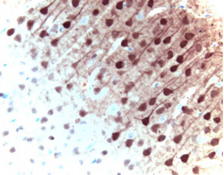
Immunohistochemical staining of ERK1 in pyrimidal cells from the rat cortex: formalin-fixed paraffin-embedded tissue, with citrate pre-treatment (40X magnification).
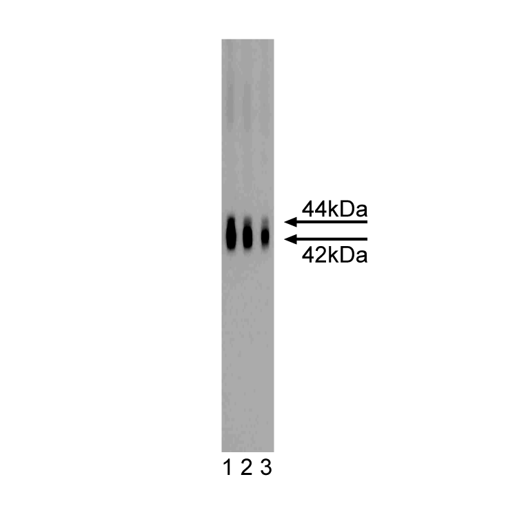
Western blot analysis of ERK1 on a rat cerebrum lysate. Lane 1: 1:4000, lane 2: 1:8000, lane 3: 1:16,000 dilution of the mouse anti-ERK1 antibody. ERK1 is expected to appear at 44 kD with crossreactivity reported to ERK2 which may appear at 42 kD.
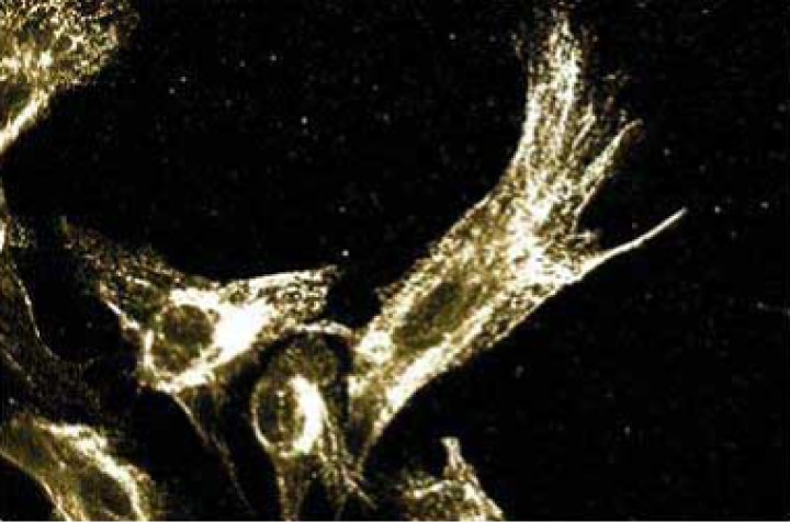
Immunofluorescence staining of human fibroblasts.

Immunohistochemical staining of ERK1 in pyrimidal cells from the rat cortex: formalin-fixed paraffin-embedded tissue, with citrate pre-treatment (40X magnification).
Western blot analysis of ERK1 on a rat cerebrum lysate. Lane 1: 1:4000, lane 2: 1:8000, lane 3: 1:16,000 dilution of the mouse anti-ERK1 antibody. ERK1 is expected to appear at 44 kD with crossreactivity reported to ERK2 which may appear at 42 kD.
Immunofluorescence staining of human fibroblasts.
Preparation And Storage
Recommended Assay Procedures
Western blot: Please refer to http://www.bdbiosciences.com/pharmingen/protocols/Western_Blotting.shtml
Product Notices
- Since applications vary, each investigator should titrate the reagent to obtain optimal results.
- Please refer to www.bdbiosciences.com/us/s/resources for technical protocols.
- Caution: Sodium azide yields highly toxic hydrazoic acid under acidic conditions. Dilute azide compounds in running water before discarding to avoid accumulation of potentially explosive deposits in plumbing.
- Source of all serum proteins is from USDA inspected abattoirs located in the United States.
Companion Products


The family of serine/threonine kinases known as ERKs (extracellular signal regulated kinases) or MAPKs (mitogen-activated protein kinases) is activated after cell stimulation by a variety of hormones and growth factors. Cell stimulation induces a signaling cascade that leads to phosphorylation of MEK (MAPK/ERK kinase) which, in turn, activates ERK via tyrosine and threonine phosphorylation. A myriad of proteins represent the downstream effectors for the active ERK and implicate it in the control of cell proliferation and differentiation, as well as regulation of the cytoskeleton. Activation of ERK is normally transient and cells possess dual specificity phosphatases that are responsible for its down-regulation. Furthermore, multiple studies have shown that elevated ERK activity is associated with some cancers. ERK1 is a 44 kDa member of the ERK family and shares 85% homology with ERK2 (42 kDa).
Development References (5)
-
Ackerley S, Grierson AJ, Brownlees J, et al. Glutamate slows axonal transport of neurofilaments in transfected neurons. J Cell Biol. 2000; 150(1):165-175. (Biology: Western blot). View Reference
-
Aguirre-Ghiso JA, Liu D, Mignatti A, Kovalski K, Ossowski L. Urokinase receptor and fibronectin regulate the ERK(MAPK) to p38(MAPK) activity ratios that determine carcinoma cell proliferation or dormancy in vivo. Mol Biol Cell. 2001; 12(4):863-879. (Biology: Western blot). View Reference
-
Nowak G. Protein kinase C-alpha and ERK1/2 mediate mitochondrial dysfunction, decreases in active Na+ transport, and cisplatin-induced apoptosis in renal cells. J Biol Chem. 2002; 277(45):43377-43388. (Biology: Western blot). View Reference
-
Reszka AA, Seger R, Diltz CD, Krebs EG, Fischer EH. Association of mitogen-activated protein kinase with the microtubule cytoskeleton. Proc Natl Acad Sci U S A. 1995; 92(19):8881-8885. (Biology: Immunofluorescence, Western blot). View Reference
-
Wan Y, Huang XY. Analysis of the Gs/mitogen-activated protein kinase pathway in mutant S49 cells. J Biol Chem. 1998; 273(23):14533-14537. (Biology: Immunoprecipitation). View Reference
Please refer to Support Documents for Quality Certificates
Global - Refer to manufacturer's instructions for use and related User Manuals and Technical data sheets before using this products as described
Comparisons, where applicable, are made against older BD Technology, manual methods or are general performance claims. Comparisons are not made against non-BD technologies, unless otherwise noted.
For Research Use Only. Not for use in diagnostic or therapeutic procedures.